Case Report 10 – Scleral RGP for Corneal Hyperalgesia
Scleral RGP for Corneal Hyperalgesia
Corneal Neuropathic Pain (Corneal Hyperalgesia)
- Ocular pain or discomfort where the pain is in excess of all clinical findings
Question is ….. How do you fit this patient if you can’t open their eyes to take any corneal topography, curvature or sag measurements?
Assumption is that these eyes are normal eyes in terms of ocular dimensions and surface profile. Fit empirically from trial set starting steeper than anticipated working backwards to anterior corneal surface with Sag calculations in microns.
- OD: 8.00/12.50/14.50/18.50/+2.25 (ORx +1.75) Sag 5600 clearance 721um
- OS: 8.00/12.50/14.50/18.50/+2.25 (ORx +1.75) Sag 5600 clearance 721um
- Ct 250um
- Clearance 100-150um for Lacosamide gtt + Lens Plus
FINAL LENS PARAMETERS
- OD: 9.00/12.50/14.25/18.50/+4.25 Sag 4999um
- OS: 9.00/12.50/14.25/18.50/+4.25 Sag 4999um
- Ct 300um Clearance 120um for Lacosamide gtt + Lens Plus
Case Report 10 – Corneal Neuropathic Pain: Scleral Lens Therapy, Hyperalgesia and Peripheral vs Central Sensitisation
Clinical Problem
This historical case demonstrates one of the more challenging areas of contemporary ocular-surface practice:
Corneal Neuropathic Pain – CNP
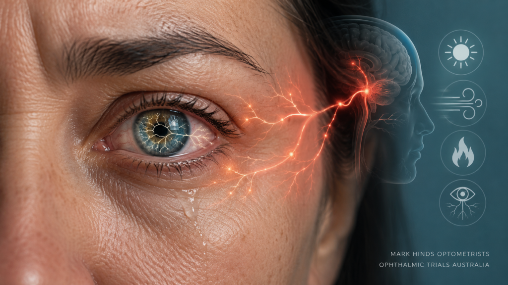
The original page described the condition as:
“Corneal hyperalgesia – ocular pain or discomfort where the pain is in excess of all clinical findings.”
That remains a useful clinical clue, but the terminology requires refinement in 2026.
A more accurate description is:
Ocular pain arising from abnormal activity within the corneal somatosensory system, often with pain severity that is disproportionate to conventional ocular-surface signs.
Importantly:
Pain disproportionate to signs is suggestive of neuropathic pain – it is not diagnostic of it.
Corneal neuropathic pain remains principally a clinical diagnosis of exclusion, and there is currently no single gold-standard diagnostic test. [1–4]
Hyperalgesia, Allodynia and Neuropathic Pain Are Not the Same Thing
Students should understand the terminology.
Hyperalgesia
An exaggerated painful response to a stimulus that would normally be painful.
For example:
a small corneal stimulus produces much greater pain than expected.
Allodynia
Pain generated by a stimulus that would not normally be painful.
Examples may include:
- normal blinking;
- mild airflow;
- wind;
- air-conditioning;
- instillation of an otherwise innocuous eye drop;
- touching the periocular region; or
- sometimes simply attempting to examine the eye.
Photoallodynia
Pain or severe discomfort triggered by light.
Spontaneous Pain
Pain occurring without an obvious external stimulus.
Patients may describe:
- burning;
- stabbing;
- aching;
- electric sensations;
- pressure;
- grittiness;
- dryness;
- foreign-body sensation;
- photophobia; or
- an inability to tolerate airflow.
These symptoms may coexist.
Teaching Point
Corneal hyperalgesia is a phenotype within abnormal nociception. Corneal neuropathic pain is the broader clinical disorder.
“Pain Without Stain” – Useful but Potentially Misleading
Corneal neuropathic pain has frequently been described as:
“Pain without stain.”
This phrase is memorable because some patients report severe pain despite:
- minimal corneal staining;
- apparently adequate tear volume;
- relatively normal slit-lamp examination; and
- no obvious structural explanation for the severity of symptoms.
However, students should not interpret this too literally.
CNP and Dry Eye Disease Can Coexist
A patient may simultaneously have:
- meibomian gland dysfunction;
- aqueous-deficient dry eye;
- tear-film instability;
- ocular-surface inflammation;
and
- peripheral or central neuropathic pain.
The presence of staining does not exclude CNP.
Likewise, absence of staining does not prove CNP.
TFOS DEWS III – Why This Case Is Even More Relevant in 2026
The TFOS DEWS III Diagnostic Methodology report substantially updated the conceptual framework for dry eye disease.
The current definition explicitly recognises:
neurosensory abnormalities
as one of the etiological factors contributing to dry eye disease. [2]
This is clinically important.
The historical division between:
“dry eye”
and
“neuropathic pain”
is often too simplistic.
Modern ocular-surface assessment needs to ask:
- Is tear-film or ocular-surface homeostasis abnormal?
- Is the pain proportional to those abnormalities?
- Is there evidence of altered corneal nociception?
- Is the pain predominantly peripheral, central or mixed?
Peripheral Versus Central Sensitisation
This distinction is fundamental to understanding why a scleral lens may help one patient dramatically and do very little for another.
Peripheral Sensitisation
Damage or inflammation affecting peripheral corneal nerves can lower nociceptor thresholds.
The peripheral nerve begins responding excessively to stimuli.
These patients may experience:
- wind sensitivity;
- pain following blinking;
- burning;
- evaporation-induced pain;
- contact sensitivity;
- photophobia; and
- exaggerated responses to ocular-surface stimulation.
A scleral lens may be particularly useful in selected patients with a substantial peripheral pain generator because it shields the corneal surface from environmental stimulation.
Central Sensitisation
Persistent peripheral nociceptive signalling can result in amplification within:
- trigeminal pathways;
- brainstem nuclei;
- thalamic pathways; and
- higher-order pain-processing networks.
Pain can then continue even when the original peripheral stimulus has been reduced.
Clinical clues may include:
- pain extending beyond the eye;
- severe photoallodynia;
- pain triggered by non-ocular stimuli;
- persistent pain despite topical anaesthesia;
- coexisting chronic pain syndromes; and
- poor correlation between ocular-surface intervention and symptoms.
Teaching Principle
A scleral lens can shield the cornea. It cannot directly switch off an established central pain network.
The Topical Anaesthetic Challenge
One of the most useful clinical tools remains the topical anaesthetic challenge.
After establishing baseline pain, a topical corneal anaesthetic such as proparacaine is instilled and the pain response is reassessed.
Complete or Near-Complete Relief
Suggests a substantial:
peripheral nociceptive / peripheral neuropathic component
because anaesthetising the peripheral corneal nerves largely removes the pain generator.
Partial Relief
Suggests:
mixed peripheral + central sensitisation
Little or No Relief
Raises concern for:
predominantly central sensitisation
or another non-corneal pain generator.
This approach is specifically incorporated into contemporary CNP assessment and TFOS DEWS III diagnostic methodology. [1–3]
But Do Not Overinterpret It
The anaesthetic challenge is a clinical localisation tool, not an infallible diagnostic test.
It does not by itself prove:
- CNP;
- absence of DED;
- a particular nerve lesion; or
- that one treatment will succeed.
The Historical Clinical Challenge
The original case asks an excellent question:
“How do you fit this patient if you can’t open their eyes to take any corneal topography, curvature or sag measurements?”
That is the defining feature of this case.
The patient’s ocular sensitivity was apparently so extreme that normal examination and imaging procedures could not initially be performed reliably.
In 2026 the ideal approach would be to obtain as much objective information as the patient can tolerate, potentially including:
- low-illumination slit-lamp examination;
- non-invasive tear-film assessment;
- anterior-segment OCT;
- corneal topography or tomography;
- corneo-scleral profilometry;
- corneal esthesiometry; and
- in vivo confocal microscopy where available.
But sometimes:
you cannot measure what the patient cannot tolerate.
That does not mean that treatment necessarily has to stop.
Historical Empirical Fitting Strategy
The historical approach was to assume that the eyes were likely to have relatively normal gross ocular dimensions and to begin with a diagnostic scleral lens providing considerably more sagittal depth than ultimately anticipated.
The fitting strategy was essentially:
Start safely above the cornea → measure what you can → work backwards in sag.
This provides an excellent practical demonstration of sagittal-depth fitting.
Initial Diagnostic Lenses
Right Eye
OD:
8.00 / 12.50 / 14.50 / 18.50 / +2.25 D
Over-refraction: +1.75 D
Sagittal depth: 5600 μm
Central clearance: approximately 721 μm
Left Eye
OS:
8.00 / 12.50 / 14.50 / 18.50 / +2.25 D
Over-refraction: +1.75 D
Sagittal depth: 5600 μm
Central clearance: approximately 721 μm
Lens Centre Thickness
250 μm
This was intentionally a very high-clearance diagnostic relationship.
It was not the intended final fit.
721 μm – Much Too Much for the Final Lens
A clearance of:
721 μm
is not an appropriate contemporary final target in an otherwise normal cornea.
A very thick post-lens reservoir:
- increases the diffusion distance for oxygen;
- may increase corneal oedema;
- increases lens mass;
- may alter centration;
- can contribute to midday fogging;
- can affect visual quality; and
- is physiologically unnecessary if much less clearance will adequately vault the cornea.
The purpose of the initial high sag in this case was different:
Avoid touching an exquisitely sensitive cornea while establishing the basic ocular geometry.
That is a defensible diagnostic strategy.
It should not be confused with a good final fit.
The Beautiful Calculation in This Case
The original diagnostic lens had:
Sag = 5600 μm
and produced:
Clearance = 721 μm
The final lens had:
Sag = 4999 μm
Difference:
5600 − 4999 = 601 μm
The final clearance was approximately:
120 μm
And:
721 − 120 = 601 μm
In other words, in this particular fitting:
a 601 μm reduction in total lens sag produced approximately a 601 μm reduction in observed central clearance.
That is an exceptionally clean teaching example.
However
Do not turn this into the rule:
“Change sag by X and clearance always changes by exactly X.”
In real scleral fitting, the relationship can be modified by:
- lens settling;
- landing-zone geometry;
- scleral compression;
- diameter;
- limbal geometry;
- lens flexure;
- decentration; and
- where the clearance is actually measured.
But in this particular historical case the relationship was remarkably close to 1:1.
Final Lens Parameters
Right Eye
OD:
9.00 / 12.50 / 14.25 / 18.50 / +4.25 D
Sagittal depth: 4999 μm
Left Eye
OS:
9.00 / 12.50 / 14.25 / 18.50 / +4.25 D
Sagittal depth: 4999 μm
Centre Thickness
300 μm
Final Clearance
Approximately:
120 μm
The final fitting therefore reduced an excessive diagnostic reservoir of approximately 721 μm to a much more conservative approximately 120 μm.
Why Is the Final Power +4.25 D Rather Than +4.00 D?
This is another useful calculation.
The initial trial lens was:
+2.25 D
with an over-refraction of:
+1.75 D
A novice might therefore expect:
+2.25 + +1.75 = +4.00 D
Yet the final lens was:
+4.25 D
Why?
Because the posterior lens geometry also changed substantially:
8.00 → 9.00 mm
and
Sag 5600 → 4999 μm
Changing posterior geometry alters the fluid-lens optical contribution.
The final lens power therefore cannot simply be calculated as:
trial lens power + original over-refraction
when the geometry of the lens has subsequently changed.
GP / Scleral Principle
Fit the geometry first. Re-over-refract the final geometry.
Right Eye – Final Scleral Lens
Figure 1. Original right-eye photograph from Case Report 10 demonstrating the final scleral lens on eye.
Left Eye – Final Scleral Lens
Figure 2. Original left-eye photograph from Case Report 10 demonstrating the final scleral lens.
Why Can a Scleral Lens Reduce Corneal Pain?
For the appropriate phenotype, the lens effectively creates a:
liquid corneal bandage
The cornea is separated from:
- eyelid friction;
- direct airflow;
- wind;
- evaporation;
- environmental particulate exposure; and
- repeated changes in the exposed tear film.
The fluid reservoir can maintain a relatively stable environment over the corneal epithelium.
Potential mechanisms for symptomatic improvement include:
- reduced stimulation of sensitised corneal nociceptors;
- reduced evaporative stress;
- reduced blink-related mechanical stimulation;
- improved epithelial hydration;
- reduced exposure to environmental triggers; and
- improvement in coexisting ocular-surface disease.
Modern scleral/PROSE literature continues to recognise neuropathic corneal pain as a potential therapeutic indication in selected patients. [8]
But a Scleral Lens Is Not an Analgesic Drug
This distinction is critical.
The lens does not directly:
- block central pain pathways;
- reverse central sensitisation;
- regenerate all abnormal corneal nerves;
- cure neuropathic pain; or
- guarantee long-term symptom relief.
It alters the peripheral environment around the cornea.
Therefore the best responders are logically more likely to be patients in whom external stimulation of peripheral corneal nociceptors remains an important pain driver.
The Paradox – Some Patients With CNP Cannot Tolerate a Scleral Lens
This is one of the most important lessons for students.
A scleral lens can dramatically reduce pain in some patients.
In another patient with severe allodynia:
the lens itself may become the painful stimulus.
Insertion can involve:
- lid manipulation;
- conjunctival contact;
- pressure from the landing zone;
- fluid touching the cornea;
- removal manipulation; and
- awareness of the lens on the ocular surface.
Patients with severe hyperalgesia or central sensitisation may find this intolerable.
Therefore:
CNP is not automatically an indication for a scleral lens.
It is an indication to consider whether peripheral shielding may help that particular pain phenotype.
What About Lacosamide?
The historical treatment recorded on this page was:
“Lacosamide gtt + Lens Plus.”
This needs an important 2026 qualification.
Lacosamide Is Not Standard Licensed Scleral-Lens Filling Therapy
Lacosamide is a sodium-channel-modulating drug primarily known as a systemic anticonvulsant.
Topical lacosamide has been proposed as an emerging/off-label treatment for peripheral corneal neuropathic pain, based on its potential to reduce abnormal nociceptor activity. [7]
However:
Evidence remains limited.
There is not yet high-quality evidence establishing topical lacosamide as routine standard therapy for CNP.
A 2024 systematic review examining therapeutic interventions for corneal neuropathy found that the overall evidence base remains limited, heterogeneous and generally of low or very-low certainty. [4]
Therefore the lacosamide used in this historical case should be presented as:
an individualised, off-label treatment used in the historical management of this patient
rather than:
a standard ingredient to place in a scleral lens reservoir.
Medication in a Scleral Lens Reservoir – Be Very Careful
A scleral lens creates prolonged exposure of the corneal surface to whatever is placed in its reservoir.
That creates potential therapeutic opportunities — but also potential risks.
Any drug placed beneath a scleral lens potentially has altered:
- residence time;
- concentration at the ocular surface;
- clearance;
- epithelial exposure; and
- pharmacokinetics.
Therefore medication in the reservoir should only be used when specifically prescribed and managed by clinicians familiar with both:
the drug
and
scleral-lens physiology.
Questions include:
- Is the formulation sterile?
- Is it preservative-free?
- What is the pH?
- What is the osmolality?
- Is it compatible with the lens material or surface coating?
- What concentration reaches the epithelium?
- How long is it retained beneath the lens?
- Is there epithelial toxicity?
- Is the treatment evidence-based?
Lens Plus – Another Historical Point
The original case documents Lens Plus as part of the filling regimen.
For a 2026 teaching page I would not recommend that students simply reproduce this historical protocol.
The contemporary default scleral-lens reservoir should generally be:
sterile preservative-free filling saline
appropriate for scleral-lens application.
A patient with corneal hyperalgesia may be particularly sensitive to preservatives, pH differences and solution composition.
Historical case regimen ≠ current universal recommendation.
The Diagnostic Work-Up in 2026
If this patient presented today, I would approach the case systematically.
1. Quantify the Pain
Document:
- intensity;
- quality;
- frequency;
- duration;
- spontaneous versus stimulus-evoked pain;
- photophobia;
- wind sensitivity;
- blink sensitivity;
- periocular pain;
- headache association; and
- impact on daily function.
A simple 0–10 numeric rating scale can be useful longitudinally.
Validated tools such as the:
- Ocular Pain Assessment Survey – OPAS
- Neuropathic Pain Symptom Inventory – Eye – NPSI-Eye
may help phenotype and monitor symptoms.
2. Examine the Ocular Surface Properly
Do not jump from:
“Lots of pain, little staining”
straight to:
“neuropathic pain.”
Assess:
- fluorescein staining;
- lissamine-green staining;
- tear-film breakup;
- tear volume;
- osmolarity where useful;
- meibomian glands;
- lid margins;
- blink;
- lagophthalmos;
- conjunctiva;
- recurrent erosion;
- epithelial basement membrane abnormalities;
- contact-lens trauma;
- infection;
- inflammation; and
- previous ocular surgery.
TFOS DEWS III specifically emphasises structured evaluation of tear-film, eyelid, ocular-surface and neural drivers. [2]
3. Exclude Other Painful Eye Disease
The differential can include:
- severe DED;
- recurrent corneal erosion;
- infectious keratitis;
- herpetic disease;
- scleritis;
- anterior uveitis;
- angle-closure mechanisms;
- post-surgical pain;
- trigeminal neuralgia;
- migraine and headache syndromes;
- referred orbital/facial pain; and
- other neurological causes.
The phrase:
“pain without stain”
should never replace a proper differential diagnosis.
4. Perform a Topical Anaesthetic Challenge
Where clinically appropriate.
This helps estimate the balance between:
peripheral
and
central
pain mechanisms.
5. Corneal Esthesiometry
If available, quantify corneal sensitivity.
Options include:
- Cochet–Bonnet esthesiometry; and
- non-contact air-pulse esthesiometry.
However, interpretation is not straightforward.
Patients with CNP can demonstrate:
- increased sensitivity;
- reduced sensitivity; or
- regional variation.
Therefore esthesiometry can support the phenotype but does not establish the diagnosis by itself. [1,2]
6. In Vivo Confocal Microscopy – IVCM
IVCM can image the sub-basal corneal nerve plexus.
Reported findings in CNP include:
- reduced nerve density;
- increased tortuosity;
- nerve beading;
- abnormal branching;
- dendritic-cell changes;
- nerve sprouts; and
- microneuroma-like structures.
A 2024 study by Liu and colleagues identified differences in clinical manifestations, corneal nerve imaging and tear neuromediator/proteomic profiles between patients with neuropathic corneal pain and controls. [5]
However:
There is currently no IVCM finding that independently proves CNP.
Some abnormalities occur in:
- dry eye;
- diabetes;
- previous surgery;
- inflammatory ocular-surface disease; and
- even some otherwise normal individuals.
IVCM should therefore support clinical reasoning rather than replace it.
7. Ask About Previous Ocular Surgery
Corneal nerve injury can occur following:
- LASIK;
- PRK;
- SMILE;
- cataract surgery;
- corneal transplantation;
- corneal infection;
- trauma; and
- other anterior-segment procedures.
Most patients heal without chronic neuropathic pain.
A small subgroup develops persistent abnormal sensory signalling.
8. Consider Systemic Pain Phenotype
CNP does not always exist in isolation.
Ask about relevant history of:
- migraine;
- facial pain;
- fibromyalgia;
- chronic widespread pain;
- trigeminal disorders;
- autoimmune disease;
- previous herpes zoster;
- neurological disease; and
- other chronic pain syndromes.
This does not mean the ocular pain is “not ocular”.
It means that the patient’s somatosensory system needs to be considered as a whole.
Multidisciplinary Management
A 2025 international survey of corneal specialists demonstrates how heterogeneous current practice remains.
Among 51 responding specialists:
- topical anaesthetic challenge was one of the most commonly used investigations;
- 69% used ocular-surface or pain questionnaires;
- IVCM was routinely used by 37%;
- blood-derived tears were commonly used;
- topical anti-inflammatory therapies were frequently employed; and
- only 38% felt comfortable independently prescribing systemic neuropathic pharmacotherapy.
Nearly half used a multidisciplinary approach, frequently involving:
- pain medicine; and
- neurology. [3]
This reinforces an important message:
Established central sensitisation is not simply an eye-drop problem.
Treatment Is Phenotype-Driven
There is no single evidence-based medication sequence that works for every patient.
Depending on the clinical phenotype, management may include combinations of:
Ocular-Surface Rehabilitation
- preservative-free lubrication;
- management of MGD;
- treatment of inflammation;
- management of allergy;
- punctal strategies where appropriate;
- autologous serum or other blood-derived tears;
- scleral/PROSE devices in selected patients.
Peripheral Neuropathic Strategies
Selected specialist-directed topical approaches may include:
- blood-derived tears;
- anti-inflammatory therapy;
- and emerging/off-label neuro-modulating strategies.
Central / Mixed Pain Strategies
May require collaboration with:
- pain specialists;
- neurologists;
- general practitioners;
- psychologists or other clinicians experienced in chronic pain;
and use of systemic neuromodulatory therapy where clinically appropriate.
Teaching Point
Treat the mechanism rather than endlessly escalating dry-eye drops because the patient says the eye feels dry.
Current Clinical Research – Ophthalmic Trials Australia & Pelthos CT2000
This is also an active area of clinical research in Brisbane.
Ophthalmic Trials Australia (OTA) is currently conducting an ocular surface pain study evaluating an investigational topical ophthalmic treatment in participants with moderate-to-severe dry eye disease and chronic ocular pain.
The study is a Phase 1b/2a clinical trial of CT2000, an investigational ophthalmic formulation of CC8464 targeting the voltage-gated sodium channel NaV1.7, which is involved in peripheral pain signalling.
Pelthos Therapeutics announced the first participant dosed in the Phase 1b/2a program on 31 March 2026. The trial includes an ascending-dose Phase 1 component and a Phase 2a component evaluating maximum tolerated dose and analgesic effect during a 28-day treatment period.
Importantly, this trial is not specifically a corneal neuropathic pain trial. Participants are being studied in the setting of moderate-to-severe dry eye disease with chronic ocular pain. It is nevertheless highly relevant to this discussion because it investigates whether selectively modulating a peripheral corneal pain pathway can reduce ocular pain without relying on conventional corticosteroid, NSAID or local-anaesthetic strategies.
For postgraduate students, this provides a useful translational link between the neurobiology discussed above and an investigational therapy currently being studied in humans.
Follow the Current Study
Ophthalmic Trials Australia – Ocular Surface Pain Study / Now Recruiting:
https://ot-au.com/now-recruiting/
Ophthalmic Trials Australia:
https://ot-au.com/
Pelthos Therapeutics – CT2000 Phase 1b/2a Eye Pain Clinical Trial:
https://pelthos.com/pelthos-therapeutics-announces-first-patient-dosed-in-phase-1b-2a-clinical-trial-of-ct2000-in-eye-pain/
Teaching Point
Corneal pain research is moving from symptom-based treatment toward mechanism-based therapy.
CT2000 is a good contemporary example: rather than simply lubricating the ocular surface, it is designed to target NaV1.7-mediated peripheral nociceptive signalling. Whether this ultimately translates into a safe and clinically useful treatment will depend on the results of controlled clinical trials.
Therapeutic Evidence – Be Humble
This is a field where clinical enthusiasm can easily move ahead of the evidence.
A 2024 systematic review by Rajan, Makrai, Lee, Singh, Chinnery and Downie found major limitations in the therapeutic evidence for corneal neuropathy.
Problems included:
- inconsistent diagnostic definitions;
- small study populations;
- heterogeneous outcome measures;
- limited randomised controlled trials; and
- low or very-low certainty for several interventions. [4]
Therefore postgraduate students should distinguish:
Biologically plausible
from
supported by case series
from
supported by controlled clinical trials.
They are not interchangeable levels of evidence.
Scleral Lens Fitting Priorities in a Pain Patient
Once a scleral lens is tolerated, fitting remains physiological.
Do not allow the pain diagnosis to justify a poor lens fit.
Assess:
- complete corneal clearance;
- limbal clearance;
- settled reservoir depth;
- landing-zone alignment;
- conjunctival compression;
- edge lift;
- lens movement;
- bubbles;
- lens decentration;
- corneal oedema;
- front-surface wetting;
- midday fogging; and
- post-removal staining.
Minimal Mechanical Stimulation Matters
A patient with corneal allodynia may respond to extremely small mechanical changes.
Pay particular attention to:
- landing-zone pressure;
- edge interaction;
- conjunctival prolapse;
- lens awareness;
- insertion trauma;
- removal trauma; and
- bubble formation.
A fit that would be considered:
“pretty good”
in an asymptomatic keratoconus patient may be completely intolerable in a patient with severe ocular allodynia.
Do Not Over-Vault to “Keep the Cornea Safe”
The initial 721 μm diagnostic clearance was useful for safely establishing geometry.
It should not become the final fit.
The final approximately:
120 μm
clearance demonstrates the appropriate principle:
reduce the reservoir once corneal safety has been established.
The goal is not maximum separation.
It is:
adequate settled clearance with minimal physiological and mechanical burden.
The Treatment Endpoint Is Pain – Not Visual Acuity
This case is fundamentally different from the previous keratoconus scleral-lens cases.
In those cases the primary endpoint was:
visual rehabilitation.
Here the principal endpoint is:
symptom reduction and functional recovery.
Relevant outcomes may include:
- baseline pain;
- end-of-day pain;
- wind tolerance;
- photophobia;
- ability to keep the eyes open;
- screen tolerance;
- driving;
- ability to work;
- time outdoors;
- sleep;
- frequency of pain exacerbations;
- wearing time;
- lens tolerance; and
- quality of life.
A patient may see:
6/6 before and after treatment
and still have achieved a life-changing therapeutic outcome.
What Would I Document Before and After the Scleral Lens?
A useful postgraduate protocol would include:
Before Lens Application
Pain NRS: /10
Photoallodynia: present/absent
Wind sensitivity: present/absent
Blink-related pain: present/absent
Corneal staining: grade
Conjunctival staining: grade
TBUT/NIBUT:
Tear volume:
Meibomian gland assessment:
Proparacaine challenge: percentage relief
Corneal sensation: where measurable
After Lens Application
At:
- 10–20 minutes;
- 1–2 hours; and
- several hours of wear where feasible,
record:
Pain NRS: /10
Photophobia: change
Wind tolerance: change
Functional improvement:
Lens awareness:
Clearance:
Landing-zone response:
Corneal physiology:
This turns:
“She seems better with the scleral lens”
into measurable clinical data.
A Particularly Useful Experimental Question
Imagine baseline pain is:
9/10
Topical anaesthetic reduces it to:
2/10
and application of a well-fitted scleral lens keeps pain around:
2–3/10
during environmental exposure.
That pattern strongly suggests that shielding the peripheral corneal pain generator is clinically meaningful.
Now consider another patient:
Baseline:
9/10
After topical anaesthetic:
8/10
After scleral lens:
8/10
The second patient has a very different phenotype.
Continuing to alter:
- sagittal depth;
- landing zone;
- filling solution; and
- lens diameter
may not solve a predominantly central pain process.
Clinical Take-Home Points
- Corneal hyperalgesia is not synonymous with corneal neuropathic pain. Hyperalgesia is an exaggerated response to a painful stimulus.
- Allodynia means pain from a normally non-painful stimulus.
- “Pain without stain” is a clue, not a diagnosis.
- CNP and genuine dry eye disease can coexist.
- TFOS DEWS III explicitly recognises neurosensory abnormalities within contemporary dry-eye pathophysiology.
- Separate peripheral, mixed and central pain mechanisms wherever possible.
- The topical anaesthetic challenge is a useful localisation tool, not a standalone diagnostic test.
- A scleral lens is most logically useful when peripheral corneal stimulation remains an important pain generator.
- A scleral lens may itself be intolerable in severe allodynia.
- The initial 721 μm clearance in this case was diagnostic — not therapeutic.
- Reducing sag from 5600 to 4999 μm reduced clearance from approximately 721 to 120 μm: a remarkable 601 μm / 601 μm relationship in this individual fit.
- Do not assume sagittal-depth changes always translate 1:1 into clearance changes.
- Changing lens geometry changes tear-lens power. Therefore the final contact-lens power cannot always be obtained by simply adding the initial over-refraction.
- The historical topical lacosamide treatment should be labelled off-label/emerging, not standard therapy.
- Do not routinely place medications into a scleral reservoir without specific clinical justification and consideration of sterility, formulation and pharmacokinetics.
- The contemporary default scleral filling solution is sterile, preservative-free saline rather than an arbitrary historical preserved saline product.
- IVCM can support a diagnosis but cannot currently prove CNP.
- Corneal sensitivity may be increased or decreased in neuropathic disease.
- Central sensitisation frequently requires multidisciplinary management.
- The primary endpoint in this case is pain and function — not Snellen acuity.
- Quantify pain before and after intervention whenever possible.
- Do not keep escalating dry-eye treatment indefinitely when the clinical phenotype suggests altered nociception.
Peer-Reviewed Reading – QUT Master of Optometry
1. Watson SL, Le DTM. Corneal neuropathic pain: a review to inform clinical practice. Eye. 2024;38(12):2350–2358. doi:10.1038/s41433-024-03060-x. PMID: 38627548.
2. Wolffsohn JS, Benítez-Del-Castillo JM, Loya-Garcia D, et al. TFOS DEWS III: Diagnostic Methodology. Am J Ophthalmol. 2025;279:387–450. doi:10.1016/j.ajo.2025.05.033. PMID: 40451408.
3. El Omda S, Tzoumas N, Calonge M, Figueiredo F. International Survey of Current Approaches to the Management of Neuropathic Corneal Pain by Experts. Ophthalmol Ther. 2025;14(12):3035–3046. doi:10.1007/s40123-025-01242-8. PMID: 41060373.
4. Rajan R, Makrai E, Lee JH, Singh S, Chinnery HR, Downie LE. Evaluating the efficacy and safety of therapeutic interventions for corneal neuropathy: A systematic review. Ocul Surf. 2024;33:80–98. doi:10.1016/j.jtos.2024.04.004. PMID: 38688453.
5. Liu C, Lin MTY, Lee IXY, et al. Neuropathic Corneal Pain: Tear Proteomic and Neuromediator Profiles, Imaging Features, and Clinical Manifestations. Am J Ophthalmol. 2024;265:6–20. doi:10.1016/j.ajo.2024.03.015. PMID: 38521157.
6. McNally TW, Figueiredo FC. Corneal Neuropathic Pain: A Patient and Physician Perspective. Ophthalmol Ther. 2024;13(4):1041–1050. doi:10.1007/s40123-024-00897-z. PMID: 38363459.
7. Nortey J, Smith D, Seitzman GD, Gonzales JA. Topical Therapeutic Options in Corneal Neuropathic Pain. Front Pharmacol. 2022;12:769909. doi:10.3389/fphar.2021.769909. PMID: 35173607.
8. Rodriguez-Garcia A, Jimenez-Perez JC, Ruiz-Lozano RE, Bustamante-Arias A, Barcelo-Canton RH. Scleral lenses and PROSE: indications, complications, and future challenges. Med Hypothesis Discov Innov Ophthalmol. 2025;14(3):73–106. doi:10.51329/mehdiophthal1525. PMID: 41111638.
9. Asiedu K. Neurophysiology of corneal neuropathic pain and emerging pharmacotherapeutics. J Neurosci Res. 2024;102(1). doi:10.1002/jnr.25285. PMID: 38284865.
10. Goyal S, Hamrah P. Understanding Neuropathic Corneal Pain—Gaps and Current Therapeutic Approaches. Semin Ophthalmol. 2016;31(1–2):59–70. doi:10.3109/08820538.2015.1114853. PMID: 26959131.
Particularly Relevant Reading for QUT Students
Watson & Le – 2024
This is probably the single best introductory paper for this case.
Students should understand from it:
- peripheral versus central CNP;
- the anaesthetic challenge;
- why clinical signs may be minimal;
- IVCM and esthesiometry;
- and why multidisciplinary treatment can be required.
TFOS DEWS III – 2025
This is essential because it places:
neurosensory abnormality
inside the modern dry-eye framework rather than treating ocular pain as an unrelated afterthought.
Rajan et al. – 2024
This is important for teaching evidence appraisal.
There are many proposed therapies for corneal neuropathic pain.
The evidence supporting them is not equally strong.
The systematic review should stop students from confusing:
“I have heard people use this”
with
“this treatment has high-certainty clinical evidence.”
Suggested Discussion Questions for Students
Question 1
A patient reports 9/10 burning ocular pain, yet demonstrates minimal corneal staining and a relatively normal tear-film examination.
What diagnoses must be excluded before calling this corneal neuropathic pain?
Question 2
Topical proparacaine reduces pain from:
9/10 → 1/10.
What does this suggest about the likely location of the dominant pain generator, and why might a scleral lens be rational in this patient?
Question 3
Another patient remains:
9/10 → 8/10
following topical anaesthesia.
Why would repeated changes to scleral lens sag probably not be the complete answer?
Question 4
The initial diagnostic lens has:
5600 μm sag and 721 μm clearance.
The final lens has:
4999 μm sag and 120 μm clearance.
Calculate both changes and explain why this particular case is such a useful demonstration of sagittal-depth fitting.
Question 5
The initial trial lens is:
+2.25 D
with:
+1.75 D over-refraction.
Why is the final lens +4.25 D rather than simply +4.00 D?
Question 6
A patient has severe photoallodynia, minimal ocular-surface disease and no pain reduction following topical anaesthetic.
What features suggest central sensitisation and which other disciplines may need to become involved?
Question 7
The historical treatment includes topical lacosamide beneath the scleral lens.
What level of evidence supports this treatment, and what additional pharmacological and safety issues arise when a medication is retained beneath a scleral lens?
Question 8
Why might an exquisitely sensitive patient experience more, rather than less, pain when a scleral lens is first inserted?
Question 9
A patient remains 6/6 before and after scleral-lens treatment but can now tolerate air-conditioning, work at a computer for eight hours and drive without severe photophobia.
Was the treatment successful? What outcome measures should document that success?
Question 10
Explain why:
“The eye looks normal, therefore the pain cannot be coming from the eye.”
is physiologically incorrect.