Case Report 2 – Keratoconus / Pellucid Marginal Degeneration
23yo female – Dietician, very active, no BIGS
GH – unremarkable
POH – LASIK review and referred in / No Gl
FOH – G, ARMD, C
OD: +0.75 / -2.00 x 72 (6/6=)* – why the dusty 6/6?
OS: +0.75 / -2.00 x 10⁵ (6/6=)* – why the dusty 6/6?
Contact Lens Fitting
- Plan A.
- J&J One Day for Astigmatism
- OD: 8.5/14.2/+0.50/-1.75 x 70 (6/6=)
- OS: 8.5/14.2/+0.50/-1.75 x 110 (6/6=)
- Plan B. – not needed
- B&L Purevision 2 for Astigmatism
- OD: 8.5/14.2/+0.50/-1.75 x 70
- OS: 8.5/14.2/+0.50/-1.75 x 110
Case Report 2 – Early Corneal Ectasia: Keratoconus vs Pellucid Marginal Degeneration Expanded
Clinical Presentation
23-year-old female dietitian, physically active.
General health: Unremarkable.
Reason for presentation: Referred for assessment in relation to possible LASIK/refractive surgery.
Past ocular history: No significant ocular history documented.
Family ocular history: Glaucoma, age-related macular degeneration and cataract.
Subjective Refraction
OD: +0.75 / −2.00 × 72 — VA 6/6=
OS: +0.75 / −2.00 × 10⁵ — VA 6/6=
At first glance, this looks like a relatively routine compound hyperopic astigmatic prescription.
The important clinical question is:
Why is the 6/6 acuity still “dusty”?
Snellen acuity is primarily a measure of high-contrast resolution. It does not adequately describe contrast sensitivity, ghosting, monocular diplopia, glare, halos or the overall quality of the retinal image.
Corneal ectasia produces increasing amounts of higher-order aberration (HOA), particularly coma and trefoil. A conventional sphero-cylindrical refraction corrects lower-order refractive error but cannot completely neutralise these irregular optical aberrations. A patient may therefore achieve 6/6 on a high-contrast chart while still reporting that the image is not genuinely sharp. [6,7]
This distinction between visual acuity and visual quality is particularly important in early keratoconus.
Corneal Topography
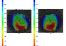
Figure 1. Corneal topography demonstrating marked inferior steepening with a crab-claw / pellucid-like configuration.
The anterior curvature maps demonstrate marked inferior steepening with a crab-claw / kissing-doves / pellucid-like configuration.
Historically, this appearance was frequently considered diagnostic of pellucid marginal degeneration (PMD). In 2026, that interpretation requires considerably more caution.
Is this actually PMD?
Not necessarily.
A crab-claw pattern on anterior corneal topography can occur in both:
- true pellucid marginal degeneration; and
- inferior or pellucid-like keratoconus.
True PMD is characterised by a peripheral, usually inferior, band of corneal thinning, classically with the area of maximal protrusion occurring superior to the zone of maximal thinning. Demonstration of the spatial relationship between thinning and protrusion is therefore more diagnostically useful than the anterior curvature pattern alone. [2–4]
2026 clinical approach
Do not diagnose PMD from the axial/sagittal curvature map alone.
Interrogate:
- anterior elevation;
- posterior elevation;
- full corneal pachymetry;
- location of the thinnest point;
- pachymetric progression;
- anterior/posterior surface asymmetry;
- epithelial thickness mapping where available; and
- corneal biomechanics where available.
Modern multimodal assessment combining tomography, OCT-derived morphology and biomechanical information improves the ability to identify early ectatic disease compared with anterior topography alone. [1,5]
For this reason, a more defensible diagnosis from the information shown is:
Bilateral corneal ectasia with an inferior/pellucid-like phenotype — keratoconus versus true PMD requiring tomographic confirmation.
The Bigger Clinical Issue: This Was a LASIK Referral
This is arguably the most important teaching point in the case.
The patient is only 23 years old and was being assessed in relation to laser refractive surgery.
Recognition of an ectatic or biomechanically susceptible cornea before tissue-removing refractive surgery is critical. Modern keratoconus assessment extends well beyond central keratometry and anterior curvature mapping, with particular attention paid to posterior elevation, pachymetric distribution and biomechanical susceptibility. [1,5]
An eye demonstrating convincing corneal ectasia should not proceed to LASIK.
Contact Lens Management
Despite the abnormal corneal shape, this patient’s refractive error remained sufficiently regular to obtain useful acuity with a conventional soft toric contact lens.
Original Fitting – Plan A
Daily disposable toric soft contact lens
OD: 8.5 / 14.2 / +0.50 / −1.75 × 70 — VA 6/6=
OS: 8.5 / 14.2 / +0.50 / −1.75 × 110 — VA 6/6=
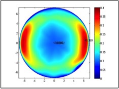
Figure 2. Thickness profile of the original daily disposable toric lens used in Plan A.
The successful fit is clinically useful because it demonstrates that a diagnosis of keratoconus does not automatically mean that a patient requires a corneal GP or scleral lens.
In mild ectasia, particularly when a substantial proportion of the astigmatism remains regular, a soft toric lens may provide satisfactory high-contrast acuity, comfort and functional vision. Specialty soft keratoconus lenses are also capable of improving high- and low-contrast visual performance in appropriately selected early-stage disease. [7,8]
But does 6/6 mean the optics are normal?
No.
A conventional soft toric lens primarily corrects sphere and cylinder. It does relatively little to neutralise significant anterior corneal irregularity and may therefore leave substantial residual HOA.
If the patient reports:
- ghosting;
- shadows around letters;
- reduced night vision;
- glare or halos;
- monocular diplopia; or
- disproportionately poor contrast despite 6/6 acuity,
then optical quality rather than Snellen acuity should drive the next stage of the contact lens fit. [6,7]
Original Fitting – Plan B
A reusable toric soft lens was retained as a second option but was not required.
OD: +0.50 / −1.75 × 70
OS: +0.50 / −1.75 × 110
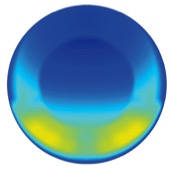
Figure 3. Thickness profile of the original reusable toric lens considered for Plan B.
2026 Teaching Note
These lens parameters and thickness-profile images should be regarded as the historical fitting data from this particular case.
They should not be interpreted as evidence that one current commercial toric lens design is universally superior for patients with ectasia. Contemporary lens availability, materials, stabilisation systems and parameter ranges change over time.
The clinical endpoint remains:
best visual quality + stable lens orientation + physiological fit + acceptable comfort.
When Would We Move Away From a Conventional Soft Toric Lens?
Escalation of the contact lens modality would be considered if spectacle or conventional soft toric correction no longer provides satisfactory visual quality.
A practical progression might include:
Conventional toric soft lens → specialty soft keratoconus lens → corneal GP / hybrid lens → scleral lens, depending on corneal geometry, disease severity, visual requirements, comfort and patient preference.
Rigid corneal and scleral lenses create a tear reservoir that masks much of the irregularity of the anterior corneal surface and can substantially reduce ocular HOAs. They do not, however, necessarily restore completely normal optics because posterior corneal and internal aberrations remain. [7]
Monitoring and Progression
At 23 years of age, diagnosis is only half of the management.
The next question is:
Is the ectasia progressing?
There is no single universally accepted parameter that should be used in isolation to define keratoconus progression.
Repeat examinations should assess changes across multiple parameters, including:
- anterior corneal curvature;
- posterior corneal shape;
- pachymetry/thinnest corneal thickness;
- refractive change;
- visual acuity;
- regional tomographic indices; and
- biomechanics where appropriate.
Kmax alone should not be regarded as the complete definition of progression. [1,9]
If reproducible progression is demonstrated, the patient should be referred for consideration of corneal cross-linking (CXL) with the primary aim of stabilising the ectatic process rather than simply improving refraction.
Clinical Take-Home Points
- 6/6 is not synonymous with normal visual quality. Patients with early ectasia may retain good high-contrast acuity while experiencing significant HOA-related degradation.
- A crab-claw topography is not synonymous with PMD. Pellucid-like keratoconus can produce a similar anterior curvature pattern.
- Look at thickness, not just curvature. The location and distribution of corneal thinning relative to the area of protrusion are important when distinguishing PMD from keratoconus.
- Tomography has superseded topography alone for ectasia assessment. OCT epithelial/stromal analysis and biomechanical assessment can provide additional information in subtle disease.
- The significance of the case is amplified by the LASIK referral. Detecting ectasia before refractive surgery may prevent iatrogenic corneal ectasia.
- Keratoconus does not automatically mean a rigid contact lens. Mild disease may function extremely well in a conventional or specialty soft toric lens.
- Treat the patient rather than the Snellen line. Contrast sensitivity, ghosting, glare and subjective visual quality may reveal optical dysfunction that 6/6 acuity misses.
- Separate visual rehabilitation from disease control. Contact lenses improve vision; they do not treat progression. Progressive disease requires consideration of CXL.
Peer-Reviewed Reading – QUT Master of Optometry
1. Gomes JAP, Hafezi F, Ambrósio R Jr, et al. Global Consensus on Keratoconus and Ectatic Diseases—Edition 2. Cornea. 2026;45(7):888–908. doi:10.1097/ICO.0000000000004170.
2. Vieira IV, Fan VH, Yu CQ. Update on pellucid marginal degeneration. Graefes Arch Clin Exp Ophthalmol. 2026;264(2):325–335. doi:10.1007/s00417-025-07022-1.
3. Tsatsos M, Koulotsiou K, Giachos I, Tsinopoulos I, Ziakas N. Pellucid marginal degeneration: a comprehensive review of pathophysiology, diagnosis, and management strategies. J Clin Med. 2025;14(15):5178. doi:10.3390/jcm14155178.
4. Lenk J, Herber R, Pillunat LE, Spörl E, Raiskup F. Differentiation between pellucid-like keratoconus and pellucid marginal degeneration using swept-source optical coherence tomography. Cornea. 2024;44(5):592–597. doi:10.1097/ICO.0000000000003648.
5. Wang X, Maeno S, Wang Y, et al. Early diagnosis of keratoconus using corneal biomechanics and OCT derived technologies. Eye Vis (Lond). 2025;12:18. doi:10.1186/s40662-025-00435-3.
6. Erdinest N, London N, Landau D, Barbara R, Barbara A, Naroo SA. Higher order aberrations in keratoconus. Int Ophthalmol. 2024;44:172. doi:10.1007/s10792-024-03118-5.
7. Swartz G, Alam K, Gentle A, Downie LE. Impact of contact lens correction on wavefront aberrations and vision quality in keratoconus. Ophthalmic Physiol Opt. 2025;45(7):1811–1828. doi:10.1111/opo.70037.
8. Gal E, Gispets J, Zyroff M, Netanya E, Gantz L. Assessment of large-diameter and small-diameter SoftK specialty contact lenses for early-stage keratoconus. Ophthalmic Physiol Opt. 2024;44(5):884–893. doi:10.1111/opo.13331.
9. Koppen C, Jiménez-García M, Kreps EO, Ní Dhubhghaill S, Rozema JJ; REDCAKE Study Group. Definitions for keratoconus progression and their impact on clinical practice. Eye Contact Lens. 2024;50(1):1–9. doi:10.1097/ICL.0000000000001038.