Case Report 4 – SynergEyes Duette HD
Case report 4 – Forme Fruste Keratoconus / High astigmatism / Irregular astigmatism
- 39yo male – American football player
- Forme Fruste K (pachs 498 um)
- OD: +4.75 / -3.00 x 1 (6/6-)
- 7.90/14.50/+4.25 medium skirt (6/6-)
- OS: +4.75 / -4.00 x 178 (6/6-)
- 7.90/14.50/+3.25 medium skirt (6/6-)
For information on the SynergEyes family of contact lenses and insertion and removal have a look here:SynergEyes Contact Lenses @ Mark Hinds Optometrists
Case Report 4 – High Astigmatism, Suspected Subclinical Keratoconus and Hybrid Contact Lenses
Clinical Presentation
39-year-old male, American football player.
The original case was recorded as “forme fruste keratoconus” with central pachymetry of approximately 498 µm, together with high hyperopic astigmatism.
Spectacle Refraction
OD: +4.75 / −3.00 × 1 — VA 6/6−
OS: +4.75 / −4.00 × 178 — VA 6/6−
This is an interesting refractive profile: significant hyperopia combined with approximately 3.00–4.00 D of predominantly with-the-rule astigmatism, yet relatively preserved high-contrast visual acuity.
The clinical question is therefore not simply:
“Can this patient read 6/6?”
It is:
“What is producing the astigmatism, how regular is the cornea, and can we improve the quality and stability of vision?”
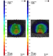
Corneal Topography
Figure 1. Original corneal topography from the case. Interpretation of possible ectasia should not be based on this anterior curvature map or pachymetry alone.
Is This Really “Forme Fruste Keratoconus”?
This is where the interpretation of the original case needs to be updated for 2026.
Historically, terms such as forme fruste keratoconus, subclinical keratoconus, keratoconus suspect and preclinical keratoconus have often been used inconsistently.
A corneal thickness of 498 µm is relatively thin, but thin does not equal keratoconus.
Central or thinnest pachymetry cannot diagnose ectasia in isolation.
Current assessment of suspected early keratoconus should integrate multiple domains, including:
- anterior corneal curvature and asymmetry;
- anterior elevation;
- posterior corneal elevation;
- thinnest pachymetry and its spatial location;
- pachymetric progression;
- epithelial thickness profile;
- inter-eye asymmetry;
- corneal biomechanics; and
- longitudinal evidence of change.
Modern tomography and biomechanical testing have substantially changed our ability to detect corneal susceptibility before obvious clinical keratoconus develops.
Consequently, based only on the information retained in this historical case, a more defensible 2026 description would be:
High bilateral corneal astigmatism with a relatively thin cornea and historical suspicion of subclinical ectasia. Further tomographic and biomechanical assessment would be required to establish or refute keratoconus.
This distinction is important.
Teaching Point
Do not diagnose keratoconus simply because the cornea is thin.
Likewise, do not rule out early ectasia simply because the patient sees 6/6.
Why Might This Patient Still Benefit From a Specialty Contact Lens?
The spectacle refraction corrects the patient’s lower-order refractive error extremely well.
However, high corneal astigmatism presents several practical contact lens problems.
A conventional toric soft contact lens must maintain its rotational position for the cylindrical correction to remain accurately aligned.
With −3.00 to −4.00 D of cylinder, relatively small amounts of lens rotation can produce appreciable residual astigmatism and variable vision.
A hybrid lens takes a different optical approach.
Hybrid Contact Lens Design
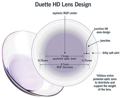
Figure 2. Original Duette HD design used in this case. The lens combines a rigid central optic with a surrounding soft skirt.
A hybrid lens combines:
a rigid gas-permeable central optic
with
a surrounding soft contact lens skirt.
The rigid central component provides the optical advantages associated with a GP lens, while the larger soft skirt assists centration, stability and subjective lens comfort.
The rigid centre means that corneal astigmatism can be substantially neutralised by the post-lens tear layer, rather than requiring the entire cylindrical correction to be incorporated into a rotating soft toric lens.
This makes hybrid lenses particularly interesting in patients with high corneal astigmatism.
Clinical research in patients with regular astigmatism greater than 3.00 D has demonstrated significant improvements in visual acuity, contrast sensitivity and glare performance with spherical hybrid lenses, with generally good patient acceptance. [4]
Original Contact Lens Fit
Right Eye
Spectacle Rx: +4.75 / −3.00 × 1 — 6/6−
Historical Duette HD:
7.90 / 14.50 / +4.25 / Medium skirt — 6/6−
Left Eye
Spectacle Rx: +4.75 / −4.00 × 178 — 6/6−
Historical Duette HD:
7.90 / 14.50 / +3.25 / Medium skirt — 6/6−
An obvious question for students is:
If the patient was already 6/6− in spectacles, why fit a hybrid lens?
Because Snellen acuity is not the complete endpoint of contact lens fitting.
With a ceiling effect around 6/6, there may be little opportunity to gain additional Snellen lines.
Potential advantages instead include:
- more stable vision;
- reduced influence of lens rotation;
- improved contrast;
- improved visual quality under mesopic conditions;
- less fluctuation following a blink;
- potentially better sports vision; and
- freedom from a high hyperopic/astigmatic spectacle correction.
For a highly active patient, those functional differences may be considerably more important than moving from 6/6− to 6/6.
Why Doesn’t the Contact Lens Power Equal the Spectacle Prescription?
This is another useful teaching component of this case.
You cannot simply transpose:
+4.75 / −4.00 × 178
into a spherical hybrid lens.
The rigid central lens creates a tear lens between its posterior surface and the anterior cornea.
The power of that tear lens depends on the relationship between:
back optic zone radius ↔ corneal curvature.
The final contact lens power therefore incorporates:
ocular refractive error + induced tear-lens power + relevant vertex-distance effects.
This is why the final powers in this case were:
OD +4.25 D
and
OS +3.25 D
rather than simply reproducing the spectacle spherical component or spherical equivalent.
This concept becomes fundamental when moving from soft contact lens fitting into corneal GP, hybrid and scleral lens optics.
How Does the Hybrid Lens Sit on the Eye?
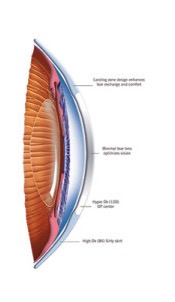
Figure 3. Cross-sectional illustration of the hybrid lens concept, demonstrating the rigid optical centre and surrounding soft skirt.
The fitting relationship has two interdependent components:
Central rigid zone: responsible predominantly for optical performance and the relationship with the corneal surface.
Soft skirt: assists lens centration, movement, comfort and overall fitting behaviour.
Students should therefore avoid assessing a hybrid lens as though it were simply either a GP or a soft lens.
It is both.
Important 2026 Product Update
The lens parameters above represent the historical Duette HD fit used in this patient.
They should not be used as a current fitting guide.
Current SynergEyes Duette technology remains a hybrid platform combining a high-Dk rigid centre with a silicone-hydrogel skirt, and contemporary versions may incorporate Tangible Hydra-PEG surface treatment.
Importantly, the current manufacturer’s positioning separates two clinical indications:
Duette — regular corneas and corneal astigmatism
UltraHealth — keratoconus and irregular corneas
This creates an important teaching issue in this historical case.
If contemporary multimodal investigation confirmed that this patient genuinely had keratoconus or significant irregular ectasia, current Duette would not be the appropriate SynergEyes design according to the manufacturer’s fitting guidance.
If, however, contemporary tomography demonstrated a structurally normal cornea with high regular corneal astigmatism, Duette remains conceptually well suited to this refractive problem.
This is why diagnosis comes before lens selection.
What Would I Measure in 2026?
If this patient presented today, I would want more than the original topography and a single pachymetry value.
The work-up should include Scheimpflug tomography assessing anterior and posterior elevation, pachymetric progression and the thinnest point.
Where available, I would add biomechanical assessment such as Corvis ST, particularly when deciding whether the cornea represents normal thin anatomy, a keratoconus suspect or true ectatic disease.
Epithelial thickness mapping can provide another layer of information because epithelial remodelling may partially mask underlying stromal irregularity.
Modern keratoconus diagnosis is increasingly multimodal rather than single-index based. [1–3]
Clinical Take-Home Points
- Thin cornea ≠ keratoconus. A pachymetry measurement of 498 µm cannot establish forme fruste keratoconus.
- High astigmatism ≠ keratoconus. Determine whether the astigmatism is regular, irregular, corneal or internal.
- 6/6 does not mean there is nothing to improve. Visual stability, contrast, glare and subjective quality may be more relevant than another Snellen letter.
- Hybrid lenses combine GP optics with a soft skirt. The rigid centre can neutralise substantial corneal astigmatism without relying on a toric soft lens maintaining an exact rotational position.
- Understand the tear lens. GP and hybrid lens power cannot simply be copied from the spectacle prescription.
- Historical lens fits should not become modern fitting guides. Product design, materials and fitting philosophy evolve.
- Use the correct lens for the correct cornea. Current Duette is directed towards regular corneal astigmatism; confirmed irregular ectasia requires an irregular-cornea design or another appropriate specialty lens modality.
- Diagnosis and visual rehabilitation are separate decisions. First determine what the cornea is doing; then select the most appropriate optical correction.
Peer-Reviewed Reading – QUT Master of Optometry
1. Gomes JAP, Hafezi F, Ambrósio R Jr, et al. Global Consensus on Keratoconus and Ectatic Diseases—Edition 2. Cornea. 2026;45(7):888–908. doi:10.1097/ICO.0000000000004170.
2. Randleman JB, Susanna BN, Hammoud B, et al. Evaluating the Global Consensus on Keratoconus and Ectatic Diseases Agreements Reached on Subclinical Keratoconus. Am J Ophthalmol. 2025;275:27–35. doi:10.1016/j.ajo.2025.03.013.
3. Wang X, Maeno S, Wang Y, et al. Early diagnosis of keratoconus using corneal biomechanics and OCT derived technologies. Eye Vis (Lond). 2025. PMID: 40350508.
4. Abou Samra WA, El-Emam DS, Kasem MA. Clinical Performance of a Spherical Hybrid Lens Design in High Regular Astigmatism. Eye Contact Lens. 2018;44(Suppl 1)–S70. doi:10.1097/ICL.0000000000000326.
5. Serdarov A, Bozkurt B, Karaküçük Y, Okudan S. Clinical Performance and Patient Satisfaction of Hybrid Contact Lenses in Patients with Keratoconus. Turk J Ophthalmol. 2023;53(4):206–212. doi:10.4274/tjo.galenos.2022.43077.
6. Ozcan SC, Ozarslan Ozcan D. Effects of a new-generation hybrid contact lens on visual performance and vision-related quality of life in patients with keratoconus. Arq Bras Oftalmol. 2023;86(1):7–12. doi:10.5935/0004-2749.20230001.
7. Çağıl N, et al. Comparison of Clinical and Topographic Outcomes of Hybrid and Scleral Lenses in Advanced Keratoconus. Turk J Ophthalmol. 2022. PMID: 35265804.
Suggested Discussion Question for Students
A 39-year-old patient has 4.00 D of refractive cylinder, 6/6− spectacle acuity and a central corneal thickness of 498 µm.
What additional evidence would you require before calling this keratoconus, and how would that diagnosis change your choice between a conventional toric soft lens, Duette hybrid, corneal GP, irregular-cornea hybrid and scleral lens?