Case Report 6 – Duette Ultrahealth SynergEyes Hybrid Contact Lenses
Duette Ultrahealth SynergEyes Hybrid Contact Lenses

- Mild K < 55D (Pachs 463 / 455um)
- Bilateral CLX with minimal ant stromal haze / prom corneal nerves / mild steepening OU
- OD: -0.75 / -4.75 x 163 (6/12+)
- Medium 200/14.5/-2.00 (6/7-) – vault 60um
- OS: -1.00 / -3.50 x 12 (6/9)
- Medium 200/14.5/-2.00 (6/7+) – vault 65um
- (OU 6/6-)
- Solution: H2O2 + Lens Plus + Hyalo Forte
Remember handy clinical signs of keratoconus at the SLIT LAMP
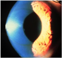
- Fleischer Ring
- Vogt’s Striae
- Stromal thinning
- Prominent Corneal Nerves – easy with dark iris
- Anterior Stromal Scars
- Sometimes due to contact lenses
- Sometimes due to CXL
- Sometimes a little bumpy on the corneal surface
- Thin cross section helps with corneal profile
For information on the SynergEyes family of contact lenses and insertion and removal have a look here:SynergEyes Contact Lenses @ Mark Hinds Optometrists
Case Report 6 – Keratoconus After Corneal Cross-Linking: SynergEyes UltraHealth Hybrid Contact Lenses
Clinical Presentation
This historical case demonstrates bilateral keratoconus treated with corneal cross-linking (CXL) and subsequently rehabilitated using SynergEyes UltraHealth hybrid contact lenses.
The original clinical findings were:
- bilateral keratoconus, historically described as “mild K <55 D”
- pachymetry approximately 463 μm OD / 455 μm OS
- previous bilateral CXL
- minimal anterior stromal haze
- prominent corneal nerves
- residual corneal steepening OU
- significant irregular refractive astigmatism
Spectacle Refraction
OD: −0.75 / −4.75 × 163 — VA 6/12+
OS: −1.00 / −3.50 × 12 — VA 6/9
This immediately gives us the key problem:
Why does conventional sphero-cylindrical correction not restore normal acuity?
Because in keratoconus the optical problem extends beyond sphere and cylinder.
Progressive corneal asymmetry generates substantial higher-order aberration, particularly coma, together with irregular astigmatism that cannot be fully corrected with spectacles.
UltraHealth Hybrid Lens Design
Figure 1. Original UltraHealth lens illustration from this case. The rigid central optic is surrounded by a silicone-hydrogel soft skirt, with a vaulted reverse-geometry fitting relationship over the irregular cornea.
First 2026 Update – This Is Not “Duette UltraHealth”
The historical page was titled:
“Duette UltraHealth SynergEyes Hybrid Contact Lenses.”
Current terminology should simply be:
SynergEyes UltraHealth
Duette and UltraHealth are different members of the SynergEyes hybrid family.
Duette is principally designed for regular corneas and corneal astigmatism.
UltraHealth is specifically designed for keratoconus and irregular corneas.
Both combine a rigid central optic with a soft peripheral skirt, but the fitting philosophies are different.
UltraHealth uses a vaulted/reverse-geometry design intended to clear the ectatic corneal apex rather than bearing directly upon it.
Historical UltraHealth Fit
Right Eye
Spectacle Rx:
−0.75 / −4.75 × 163 — 6/12+
Historical UltraHealth:
Medium skirt / 200 μm vault / 14.5 mm / −2.00 D
VA: 6/7−
Measured apical clearance: approximately 60 μm
Left Eye
Spectacle Rx:
−1.00 / −3.50 × 12 — 6/9
Historical UltraHealth:
Medium skirt / 200 μm vault / 14.5 mm / −2.00 D
VA: 6/7+
Measured apical clearance: approximately 65 μm
Binocular Outcome
OU: 6/6−
This is a substantial functional improvement.
The right eye improves from approximately:
6/12+ → 6/7−
and the left from:
6/9 → 6/7+
while binocularly achieving approximately 6/6−.
How Can a −2.00 D Spherical Hybrid Lens Correct −4.75 D of Refractive Cylinder?
This is one of the best teaching questions in the case.
The spectacle prescription in the right eye contains:
−4.75 D cylinder
yet the successful hybrid lens power is simply:
−2.00 D sphere.
How?
Because the rigid optical component changes the optical system.
A rigid anterior lens surface creates a much more regular refracting surface and the fluid layer between the posterior lens and irregular anterior cornea substantially masks the anterior corneal irregularity.
The spectacle prescription is trying to correct the total ocular aberration using:
sphere + cylinder
whereas the hybrid lens is effectively replacing much of the irregular anterior corneal refracting surface with a regular rigid optical surface.
Consequently:
A spherical GP or hybrid lens can correct large amounts of corneal astigmatism and irregularity without incorporating the same cylindrical power found in the spectacle prescription.
This is a fundamental difference between:
soft toric optics
and
rigid optics.
Why Doesn’t the Final Lens Power Follow the Spherical Equivalent?
Again, because this is not simply a spectacle-to-contact-lens vertex conversion.
Final UltraHealth power is influenced by:
- the patient’s refractive error;
- the irregular corneal shape;
- the posterior lens geometry;
- the fluid/tear-lens effect;
- vault;
- lens centration; and
- the final over-refraction.
The two eyes in this case have noticeably different spectacle prescriptions yet ultimately required the same −2.00 D lens power.
That is a useful demonstration of why specialty rigid lens prescribing should be driven by:
fit + over-refraction
rather than simply transposing the spectacle prescription.
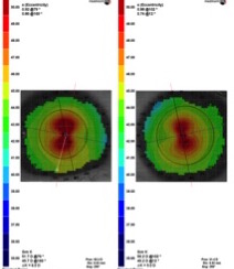
Corneal Topography
Figure 2. Original bilateral topography from the case, demonstrating asymmetric corneal steepening consistent with keratoconus.
“Mild Keratoconus Because K Is <55 D”?
This is another part of the historical page that should be updated.
Older classifications often divided keratoconus into categories according to keratometric thresholds.
That is no longer sufficient.
Kmax alone does not define keratoconus severity.
Likewise:
463 μm / 455 μm pachymetry does not by itself stage this disease.
The contemporary assessment of keratoconus should integrate multiple structural and functional parameters, including:
- anterior curvature;
- posterior elevation;
- thinnest pachymetry;
- location of the thinnest point;
- pachymetric progression;
- anterior and posterior radius of curvature;
- refractive error;
- corrected visual acuity;
- epithelial thickness where available;
- corneal biomechanics where available; and
- longitudinal change.
The 2026 Global Consensus on Keratoconus and Ectatic Diseases – Edition 2 reinforces this multimodal approach rather than reliance on a single keratometry or pachymetry number. [1]
Where appropriate, contemporary documentation might also include Belin ABCD staging rather than simply referring to “mild”, “moderate” or “severe” keratoconus on the basis of Kmax alone.
Cross-Linking and Contact Lenses Do Different Jobs
This patient had already undergone bilateral corneal cross-linking.
This creates a fundamental teaching distinction.
CXL
Purpose: biomechanical stabilisation of progressive ectasia
Contact Lens
Purpose: optical rehabilitation
The UltraHealth lens does not treat keratoconus progression.
CXL does not necessarily provide the patient with normal optics.
The two treatments therefore address different problems.
A patient can have successfully stabilised keratoconus after CXL and still have:
- irregular astigmatism;
- coma;
- ghosting;
- reduced contrast;
- reduced spectacle BCVA; and
- a continued need for specialty contact lenses.
Do Hybrid Lenses Cause Progression After CXL?
Available evidence is reassuring but should not be overstated.
A study comparing post-CXL keratoconus patients wearing UltraHealth hybrid lenses with spectacle wearers found no significant difference in topographic changes between groups over 12 months. [4]
That does not mean contact lenses stabilise keratoconus.
It means that appropriately fitted hybrid lens wear was not associated with evidence of increased progression in that study.
The 60–65 μm Vault – Why Does It Matter?
The historical OCT measurements were:
OD approximately 60 μm
OS approximately 65 μm
This was an excellent teaching feature then and remains so now.
UltraHealth is designed to vault the corneal apex.
Current SynergEyes fitting information describes a final central/apical clearance in the approximate 50–100 μm range, while current Instructions for Use specify approximately 100 μm central apical clearance after fitting.
Therefore, the historical 60–65 μm values represent a relatively low but intentional clearance relationship rather than corneal bearing.
But Be Precise About Where You Measure It
In keratoconus:
the cone apex may not correspond to the visual axis or geometric corneal centre.
If OCT clearance is measured through the wrong meridian or through the pupil centre rather than through the ectatic apex, the result may substantially misrepresent the actual minimum clearance.
2026 Assessment
Use anterior-segment OCT where available to measure:
- clearance over the true cone apex;
- minimum corneal clearance;
- rigid-soft junction relationship;
- inner landing-zone relationship; and
- peripheral/skirt behaviour.
Do not simply place one central OCT caliper through the visual axis and call that the lens clearance.
Why Not Increase the Vault to 200 or 300 μm?
Because more clearance is not automatically better.
The objective is not to create the largest possible fluid reservoir.
The objective is:
sufficient clearance to avoid clinically significant corneal bearing while maintaining appropriate movement, oxygen delivery, tear exchange, centration and comfort.
Excessive vault can alter:
- lens movement;
- lens stability;
- tear exchange;
- optical performance;
- oxygen availability; and
- overall lens behaviour.
This is a major conceptual distinction between an UltraHealth hybrid and a large-diameter scleral lens.
The hybrid lens is intended to vault the ectatic apex but continue to behave as a relatively mobile 14.5-mm hybrid lens.
The Soft Skirt Still Matters
It is tempting to focus entirely on the rigid centre.
That is a mistake.
The soft skirt contributes substantially to:
- centration;
- movement;
- comfort;
- lens retention;
- alignment; and
- tear exchange.
A suitable central vault with an inappropriate skirt is still a poor fit.
Contemporary UltraHealth assessment should therefore include:
- apical clearance;
- inner landing zone;
- skirt alignment;
- edge behaviour;
- movement on blink;
- centration;
- bubbles;
- comfort; and
- post-lens epithelial integrity.
Current manufacturer guidance expects approximately 1 mm of movement on blink, rather than a completely immobile lens.
The 2026 Evidence for UltraHealth
There is now considerably better peer-reviewed evidence for this lens than when the original case page was written.
A 2026 study specifically evaluating SynergEyes UltraHealth examined patients with moderate-to-advanced keratoconus or irregular astigmatism who had inadequate visual rehabilitation with conventional correction.
Among the eyes continuing UltraHealth wear at six months:
- mean lens-corrected acuity improved significantly;
- 88.6% achieved approximately 6/9.5 (0.22 logMAR) or better;
- NEI-VFQ-25 scores improved significantly across several domains;
- near and distance activity scores improved;
- general vision improved; and
- no serious ocular complications were observed during follow-up. [2]
This is important because contact lens success should not be assessed purely by:
“What line can they read?”
It should also consider:
- functional vision;
- comfort;
- wearing time;
- near activity;
- distance activity;
- visual confidence; and
- vision-related quality of life.
But Hybrid Lenses Are Not Successful in Everybody
Students should also see the other side of the evidence.
A 2021 clinical-outcomes study of 102 keratoconic eyes found significant improvement in visual acuity with hybrid lenses.
However, 37.8% of eyes discontinued hybrid lens wear within six months.
The major reasons were:
- discomfort;
- handling difficulties; and
- less commonly, inadequate vision.
Success also correlated with cone morphology. [3]
This is clinically important.
A lens can have excellent optics and still fail.
Successful contact lens prescribing requires:
optics + physiology + handling + comfort + patient motivation.
Hybrid Versus Scleral – Which Is Better?
There is no universal winner.
Both are excellent visual rehabilitation options for keratoconus.
A comparative study of hybrid and mini-scleral lenses in advanced keratoconus found that both significantly improved:
- visual acuity;
- refractive outcome;
- corneal higher-order aberrations; and
- functional optical performance.
However, greater comfort was reported with the mini-scleral lenses in that study. [5]
Reasons I Might Prefer an UltraHealth Hybrid
- good rigid-lens optical response;
- relatively small overall diameter;
- useful centration;
- adequate corneal vault;
- patient prefers hybrid handling;
- minimal ocular surface disease;
- satisfactory comfort;
- no need for a large scleral reservoir.
Reasons I Might Prefer a Scleral Lens
- very advanced or highly decentered cone;
- poor hybrid comfort;
- hybrid instability;
- significant ocular surface disease;
- corneal scarring/irregularity requiring complete vault;
- intolerance of corneal or hybrid designs;
- desire for greater corneal/limbal protection;
- persistent poor vision in another modality.
Teaching Point
Do not fit a scleral lens because:
“Keratoconus = scleral.”
Likewise, do not fit a hybrid simply because:
“Hybrid = more comfortable GP.”
Choose the lens based on the individual cornea and patient.
Slit-Lamp Signs of Keratoconus
Figure 3. Original slit-lamp image demonstrating a Fleischer ring associated with keratoconus.
Topography and tomography have transformed keratoconus diagnosis, but fourth-year optometry students should still be able to recognise the classic slit-lamp signs.
Fleischer Ring
A yellow-brown to green-brown ring or arc of iron deposition within the corneal epithelium, usually surrounding the base of the cone.
It may be partial rather than complete.
The ring is often easier to appreciate with appropriate slit-lamp illumination and cobalt-blue assessment.
Vogt Striae
Fine vertical or oblique stress lines within the:
- deep stroma;
- Descemet membrane; and/or
- posterior corneal region.
They reflect mechanical stress associated with ectasia.
Their presence is supportive of established keratoconus but their absence certainly does not exclude disease.
Stromal Thinning
Look at the corneal profile with a thin optic section.
Do not rely solely on an apparently normal anterior surface.
Modern tomography quantifies thickness distribution much more accurately, but the ability to recognise thinning clinically remains important.
Prominent Corneal Nerves
Corneal nerves may appear unusually prominent in keratoconus.
This is a recognised finding but is not sufficiently specific to diagnose keratoconus on its own.
Increased visibility can be especially obvious against a dark iris and in a thin cornea.
Corneal Scarring
More advanced keratoconus may produce:
- anterior stromal scarring;
- apical scarring;
- deeper stromal opacity; and
- scarring following episodes of acute hydrops.
Chronic mechanical contact-lens bearing has historically also been associated with apical epithelial/stromal trauma and scarring.
Avoid This
A specialty lens should not be deliberately fitted with substantial chronic apical bearing simply because the cornea is steep.
CXL Haze Is Not Necessarily Keratoconus Scarring
The historical notes describe minimal anterior stromal haze following CXL.
This distinction is important.
Corneal cross-linking can produce postoperative stromal haze, particularly during the healing response.
Haze should not automatically be labelled:
“keratoconus scar.”
Likewise, the presence of anterior stromal opacity after CXL does not necessarily imply:
- progressive keratoconus;
- contact-lens damage; or
- treatment failure.
Clinical interpretation should consider:
- timing since CXL;
- density and depth of opacity;
- change over time;
- BCVA;
- densitometry where available; and
- other evidence of corneal progression.
Stromal haze is a recognised complication or healing response following CXL, while true significant scarring is less common. [7,8]
Modern Slit-Lamp Signs – Useful but Not Required for Early Diagnosis
A patient with early keratoconus may have:
- no Fleischer ring;
- no obvious Vogt striae;
- no clinically obvious thinning;
- no scar; and
- apparently normal slit-lamp findings.
That does not exclude keratoconus.
Modern diagnosis increasingly occurs before classical slit-lamp signs develop, using:
- tomography;
- epithelial thickness mapping;
- posterior corneal assessment; and
- biomechanics.
This is exactly why keratoconus is now detected considerably earlier than it was when these clinical classifications were developed. [1,6]
Lens Care – Important 2026 Update
The historical case recorded:
H₂O₂ + Lens Plus + Hylo-Forte
as the solution regimen.
For teaching purposes, this should now be separated into two different concepts:
1. Disinfection
A hydrogen-peroxide system can be appropriate for UltraHealth when used exactly according to the lens and solution instructions and allowed to fully neutralise before the lens is placed on the eye.
Never place unneutralised hydrogen peroxide in the eye.
2. Lens Insertion
Current SynergEyes instructions recommend filling the UltraHealth lens bowl completely with:
preservative-free sterile saline
before insertion.
This helps:
- prevent air bubbles;
- maintain the fluid reservoir; and
- allow the vaulted lens to settle appropriately over the cornea.
Do Not Use Tap Water
Tap water should never be used to:
- rinse;
- fill;
- store; or
- wet specialty contact lenses.
This is an important microbial-keratitis and Acanthamoeba prevention message.
If a preservative-free viscous lubricant such as a hyaluronic-acid product is added to the lens reservoir for an individual patient, that should be regarded as clinician-directed use rather than simply assumed to be the manufacturer’s standard filling recommendation.
If the lens carries Tangible Hydra-PEG, care products must also be compatible with the coating.
What Would I Measure if This Patient Presented in 2026?
1. Establish Whether the Keratoconus Is Stable
Previous CXL does not eliminate the need for surveillance.
Compare serial:
- tomography;
- anterior and posterior curvature;
- pachymetric progression;
- thinnest pachymetry;
- ABCD parameters;
- refraction;
- BCVA; and
- biomechanics where appropriate.
2. Document the Corneal Surface
Look specifically for:
- Fleischer ring;
- Vogt striae;
- stromal thinning;
- apical scarring;
- CXL haze;
- epithelial disruption;
- prominent nerves; and
- signs of previous hydrops.
3. Assess Lens Clearance With OCT
Measure through the true cone apex, not simply the pupil centre.
Document:
- initial clearance;
- settled clearance;
- minimum clearance;
- relationship at the GP/soft junction; and
- any localized corneal bearing.
4. Assess Movement and Skirt Alignment
Look for:
- approximately appropriate blink movement;
- stable centration;
- no significant skirt wrinkling;
- no excessive edge lift;
- no significant compression;
- adequate tear exchange; and
- comfortable recovery following the blink.
5. Remove the Lens and Look Again
The post-removal examination can be more informative than the lens-on-eye appearance.
Assess:
- epithelial staining;
- indentation/compression;
- localized apical trauma;
- limbal hyperaemia;
- conjunctival response; and
- corneal oedema.
6. Measure More Than High-Contrast VA
This patient’s improvement to:
OD 6/7−
OS 6/7+
OU 6/6−
is excellent.
But contemporary assessment should also consider:
- contrast sensitivity;
- ghosting;
- monocular diplopia;
- night driving;
- glare;
- halo;
- wearing time;
- comfort;
- handling; and
- patient-reported functional vision.
Clinical Take-Home Points
- UltraHealth and Duette are different SynergEyes lens families. UltraHealth is the irregular-cornea/keratoconus design.
- CXL treats progression; a contact lens treats vision. Never confuse disease stabilisation with optical rehabilitation.
- Kmax <55 D does not automatically mean “mild keratoconus.” Modern staging requires more than one keratometric number.
- Pachymetry alone does not stage the disease. A cornea measuring 455 μm tells you that it is relatively thin, not how severe or progressive the keratoconus is.
- A spherical hybrid lens can correct large amounts of irregular astigmatism. The rigid central optic masks much of the anterior corneal irregularity.
- The spectacle cylinder does not need to appear in the final hybrid lens prescription.
- Measure clearance at the cone apex. Measuring OCT clearance through the visual axis can be misleading in a decentered cone.
- More vault is not automatically better. The goal is adequate clearance with appropriate movement, oxygenation, tear exchange, centration and comfort.
- The skirt is part of the fit. Do not assess only central clearance.
- Hybrid lenses can provide excellent vision but are not universally tolerated. Handling and comfort remain clinically important causes of discontinuation.
- Scleral and hybrid lenses are alternatives, not a hierarchy. Select the modality that best fits the cornea and patient.
- Recognise the classical slit-lamp signs. Fleischer ring, Vogt striae, thinning, prominent nerves and stromal scarring remain useful even in the era of advanced tomography.
- Distinguish CXL haze from keratoconus scarring or contact-lens trauma.
- Contact lens care is part of the prescription. Preservative-free sterile saline for insertion, appropriate disinfection and no tap water.
- Treat the patient’s visual function, not just the Snellen line.
Peer-Reviewed Reading – QUT Master of Optometry
1. Gomes JAP, Hafezi F, Ambrósio R Jr, et al. Global Consensus on Keratoconus and Ectatic Diseases—Edition 2. Cornea. 2026;45(7):888–908. doi:10.1097/ICO.0000000000004170. PMID: 42228627.
2. Cakmak-Cengiz E, Karaman-Atasever F, Can E. Restoring functional vision in moderate-advanced keratoconus: quality-of-life outcomes with hybrid contact lenses. Int Ophthalmol. 2026;46:157. doi:10.1007/s10792-026-04028-4. PMID: 41831166.
3. Kloeck D, Koppen C, Kreps EO. Clinical Outcome of Hybrid Contact Lenses in Keratoconus. Eye Contact Lens. 2021;47(5):283–287. doi:10.1097/ICL.0000000000000738. PMID: 33009254.
4. Aslan MG, et al. The impact of hybrid contact lenses on keratoconus progression after accelerated transepithelial corneal cross-linking. Int Ophthalmol. 2021;41(1):45–55. doi:10.1007/s10792-020-01551-w. PMID: 32856196.
5. Akcay BİS, Kockar A, Limon U, Kardes E, Dogan Dursun A. Comparison of Clinical and Topographic Outcomes of Hybrid and Scleral Lenses in Advanced Keratoconus. Beyoglu Eye J. 2022;7(1):59–65. doi:10.14744/bej.2021.29981. PMID: 35265804.
6. Niazi S, Gatzioufas Z, Doroodgar F, et al. Keratoconus: exploring fundamentals and future perspectives – a comprehensive systematic review. Ther Adv Ophthalmol. 2024;16:25158414241232258. doi:10.1177/25158414241232258. PMID: 38516169.
7. Naranjo A, Manche EE. A comprehensive review on corneal crosslinking. Taiwan J Ophthalmol. 2024;14(1):44–49. doi:10.4103/tjo.TJO-D-23-00055. PMID: 38654993.
8. Borchert GA, Kandel H, Watson SL. Epithelium-on versus epithelium-off corneal collagen crosslinking for keratoconus: a systematic review and meta-analysis. Graefes Arch Clin Exp Ophthalmol. 2024;262:1683–1692.
9. Serdarov A, Bozkurt B, Karaküçük Y, Okudan S. Clinical Performance and Patient Satisfaction of Hybrid Contact Lenses in Patients with Keratoconus. Turk J Ophthalmol. 2023;53(4):206–212.
10. Evaluation of Visual Acuity Outcomes and Corneal Alterations of New Generation Hybrid Contact Lenses in Patients With Advanced Keratoconus. Eye Contact Lens. 2020. PMID: 32732702.
Current Product-Specific Source – Not Peer Reviewed
SynergEyes. UltraHealth and UltraHealth FC Instructions for Use and professional fitting/handling resources. Current manufacturer guidance specifies approximately 100 μm central apical clearance after fitting, adequate lens movement and filling the lens with sterile preservative-free saline for insertion.
Suggested Discussion Questions for Students
Question 1
The right spectacle refraction is:
−0.75 / −4.75 × 163
yet the successful UltraHealth prescription is:
−2.00 D sphere.
Explain how a spherical rigid optical surface can produce a major improvement in vision without incorporating −4.75 D of cylinder into the lens.
Question 2
The historical OCT clearance was:
60 μm OD and 65 μm OS.
Where should this measurement be taken in a decentered keratoconic cone, and why might a measurement through the pupil centre be misleading?
Question 3
The patient has undergone bilateral CXL.
What evidence would you require before concluding that the keratoconus is now stable?
Question 4
The patient achieves excellent acuity with an UltraHealth hybrid lens.
What clinical findings might still make you refit this patient into a scleral lens?
Question 5
At slit lamp you observe mild anterior stromal opacity in an eye previously treated with CXL.
How would you distinguish CXL-related haze from apical keratoconus scarring or contact-lens-induced epithelial/stromal trauma?
Question 6
A patient has 150 μm of central clearance and excellent 6/6 acuity.
Why is it incorrect to conclude from those two findings alone that the UltraHealth fit is optimal?