Case Report 3 – Intacs / Kerarings / Keratoconus
- 27 yo male – Columbian Medical Student
- POH – NEW intracorneal ring segments OD progressive 70+D central cone / OS moderate myope
- OD: -1.75 / -1.75 x 90 (6/12)
- OS: -7.75 / -0.75 x 10⁵ (6/7)
- OD: 8.9/14.5/-1.75/-1.75X90 (6/12)
- BLPV2HDFA
- OS: 8.6/14.0/-7.50 (6/6)
- BLPV2HD
- Simple is sometimes best!
Case Report 3 – Keratoconus, Intracorneal Ring Segments and Contact Lens Rehabilitation
Clinical Presentation
27-year-old male, Colombian medical student.
The historical notes record:
- OD: progressive keratoconus with a very steep central cone (>70 D) and recent intracorneal ring segment implantation
- OS: moderate myopia with less severe corneal involvement
Spectacle Refraction
OD: −1.75 / −1.75 × 90 — VA 6/12
OS: −7.75 / −0.75 × 10⁵ — VA 6/7
Immediately, there is an important asymmetry.
The right eye has relatively modest manifest refractive error but only achieves 6/12, whereas the substantially more myopic left eye reaches approximately 6/7.
Why?
Because refractive magnitude and visual quality are not the same thing.
In keratoconus, reduced corrected acuity is driven largely by irregular corneal optics and higher-order aberrations, rather than simply the amount of sphere or cylinder measured during subjective refraction.
A patient can therefore have only modest sphero-cylindrical error but relatively poor corrected acuity if the corneal surface is sufficiently irregular.
Intracorneal Ring Segments
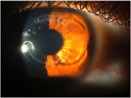
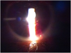
Figure 1. Original slit-lamp photograph demonstrating the intracorneal ring segment within the corneal stroma.
Intracorneal ring segments (ICRS) are polymethyl methacrylate or, increasingly, other intrastromal implants positioned within the peripheral or mid-peripheral corneal stroma.
Examples include:
- INTACS
- KeraRing
- Ferrara Ring
- other synthetic ICRS designs
- more recently, corneal allogenic intrastromal ring segments (CAIRS)
Their role is principally to alter corneal geometry.
By adding volume to the peripheral or mid-peripheral corneal stroma, the segments can produce a relative flattening and regularisation of the central cornea.
Potential outcomes include:
- reduced K values;
- reduced refractive and topographic astigmatism;
- improved corneal symmetry;
- reduced coma in selected patients;
- improved uncorrected vision;
- improved corrected vision; and
- potentially improved subsequent contact lens tolerance.
Modern outcomes depend heavily on cone morphology, segment thickness, arc length, diameter, number of segments and implantation position. [2–4]
OCT of the Ring Segment
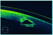
Figure 2. Anterior segment OCT demonstrating the intracorneal position of the ring segment.
Anterior segment OCT allows the clinician to assess:
- implantation depth;
- stromal position;
- relationship to the anterior and posterior corneal surfaces;
- segment position within the tunnel; and
- potential complications such as superficial placement or migration.
Contemporary implantation is commonly performed using femtosecond-laser-created stromal channels, providing greater control over tunnel geometry and implantation depth than historical manual dissection techniques.
The Critical 2026 Distinction
Does an intracorneal ring segment stop keratoconus progression?
Do not assume that it does.
This is one of the most important teaching points in this case.
ICRS and corneal cross-linking address different clinical problems:
ICRS
Primary aim: corneal reshaping and visual rehabilitation
Corneal Cross-Linking
Primary aim: biomechanical stabilisation of progressive ectasia
ICRS implantation may flatten and regularise a keratoconic cornea, but it should not be considered equivalent to CXL when progressive disease requires stabilisation.
The contemporary literature examining combined ICRS and CXL explicitly distinguishes the reshaping effect of ICRS from the biomechanical stabilisation provided by CXL. [1,5]
For a 27-year-old patient with documented progressive keratoconus, one of the first questions in 2026 should therefore be:
Has progression been demonstrated, and has the need for corneal cross-linking been addressed?
Corneal Topography
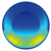
Figure 3. Original topographic images from the case illustrating the markedly different corneal geometries between the two eyes.
The historical description records a >70 D central cone OD.
At this level of steepening, simply quoting Kmax is not sufficient to describe the eye.
A contemporary keratoconus assessment would also examine:
- anterior elevation;
- posterior elevation;
- thinnest pachymetry;
- pachymetric progression;
- cone location;
- Belin ABCD staging;
- epithelial thickness profile where available;
- higher-order aberrations;
- corneal biomechanics; and
- longitudinal change.
The 2026 Global Consensus reinforces the move away from relying on a single index to diagnose, stage or assess progression in keratoconus. [1]
What Has the Ring Actually Done?
An important question for students is:
Has the cornea become normal?
No.
Intracorneal ring implantation may make the corneal shape more regular, but it does not convert an ectatic cornea into a normal cornea.
Residual abnormalities can include:
- irregular astigmatism;
- coma;
- trefoil;
- residual asymmetric power;
- reduced contrast sensitivity; and
- suboptimal spectacle-corrected acuity.
This explains why many patients continue to require contact lenses after ICRS surgery.
Contact Lens Rehabilitation After ICRS
The presence of intracorneal ring segments does not mean that contact lens wear is contraindicated.
Published studies have demonstrated successful fitting after ICRS using multiple modalities, including:
- conventional soft lenses;
- toric soft lenses;
- keratoconus-specific soft lenses;
- corneal GP lenses;
- piggyback systems;
- hybrid lenses; and
- corneo-scleral or scleral lenses. [6,7]
The correct modality is determined by the residual corneal optics and the patient’s functional requirements, not simply by the diagnosis or surgical history.
Original Contact Lens Fit – Right Eye
Spectacle Refraction
OD: −1.75 / −1.75 × 90 — VA 6/12
Soft Toric Contact Lens
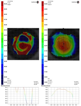
OD: 8.9 / 14.5 / −1.75 / −1.75 × 90 — VA 6/12
Figure 4. Thickness profile of the historical toric soft contact lens used for the right eye.
The right eye retained approximately 6/12 acuity.
This is clinically significant.
The soft toric lens corrects the patient’s sphere and cylinder but does relatively little to neutralise the residual irregular astigmatism and higher-order aberration associated with advanced keratoconus.
So why use it?
Because the objective of contact lens management is not automatically:
“Fit the most complicated lens possible.”
If the patient is:
- comfortable;
- functionally satisfied;
- achieving adequate binocular vision;
- physiologically tolerating the lens; and
- meeting their occupational and lifestyle requirements,
then a simple lens can sometimes be entirely appropriate.
That was the original lesson from this case:
Simple is sometimes best.
But Would We Stop at 6/12 in 2026?
Not necessarily.
For a young medical professional, I would specifically investigate whether the right eye could achieve meaningfully better visual quality with a rigid optical system.
A diagnostic trial with:
- corneal GP;
- hybrid;
- corneo-scleral; or
- scleral lens
would demonstrate whether masking the irregular anterior corneal surface improves acuity, contrast and subjective quality.
If a scleral or corneo-scleral lens produced, for example, substantially better acuity and visual quality than 6/12, the patient could then make an informed decision between:
simplicity and convenience
versus
maximum optical performance.
Published work demonstrates significant improvements in visual acuity and visual quality when corneo-scleral lenses are fitted to patients who remain visually dissatisfied after ICRS implantation. [7]
Slit-Lamp Appearance With Contact Lens
Figure 5. Original slit-lamp image demonstrating contact lens wear in the eye containing an intracorneal ring segment.
When fitting a corneal contact lens over an ICRS-treated cornea, carefully assess:
- centration;
- movement;
- corneal bearing;
- epithelial staining;
- relationship of the lens to the ring segment;
- conjunctival response;
- tear exchange;
- wearing time; and
- subjective comfort.
A poorly centred or excessively bearing rigid lens may interact unfavourably with the altered corneal geometry.
This is one reason why scleral designs can be particularly useful in selected post-ICRS eyes: the lens can vault the cornea and ring segments entirely.
Left Eye – Do Not Over-Treat the Cornea
Spectacle Refraction
OS: −7.75 / −0.75 × 10⁵ — VA 6/7
Historical Contact Lens
OS: 8.6 / 14.0 / −7.50 — VA 6/6
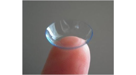
Figure 6. Conventional soft contact lens used to illustrate the simple optical solution in the less affected left eye.
Despite relatively high myopia, this eye achieved 6/6 with a simple spherical soft contact lens.
This provides a very useful contrast with the right eye.
OD
Much lower refractive error — poorer visual acuity because of irregular ectatic optics.
OS
Much greater spherical refractive error — excellent visual acuity because the optics are comparatively regular.
This reinforces a fundamental clinical principle:
The magnitude of a refractive error tells you very little about the optical regularity of the cornea.
Why Does a −7.50 D Spherical Lens Work When the Spectacle Refraction Contains Cylinder?
The left spectacle refraction contains only −0.75 D cylinder.
Several factors can allow a spherical soft lens to perform extremely well:
- some of the spectacle cylinder may be relatively small clinically;
- spherical soft lenses drape over the corneal surface;
- residual cylinder may remain below the patient’s tolerance threshold;
- binocular summation may further improve functional performance.
The measured endpoint was 6/6, so increasing lens complexity would offer little benefit unless the patient reported residual blur, glare, fluctuation or another functional complaint.
Again:
Do not make a lens more complicated simply because you can.
ICRS Complications Students Should Know
Although ICRS can provide meaningful visual and topographic improvement, these are implanted devices and are not complication-free.
Reported complications include:
- segment migration;
- extrusion;
- superficial placement;
- infectious keratitis;
- stromal thinning over the segment;
- corneal neovascularisation;
- deposits around the segment;
- glare and halos;
- persistent visual symptoms;
- incorrect positioning; and
- requirement for repositioning or explantation.
A 2026 systematic review of INTACS outcomes found consistent improvement in visual and corneal parameters but highlighted migration, extrusion and secondary surgical intervention as clinically important device-related complications. [2]
Regular postoperative surveillance therefore remains important.
Intacs, Keraring and the Changing Landscape
The original case focused on synthetic ICRS such as INTACS and KeraRing.
The field has evolved considerably.
Current options include different:
- segment diameters;
- thicknesses;
- arc lengths;
- cross-sectional profiles;
- implantation patterns; and
- patient-specific nomograms.
CAIRS – The Major Contemporary Development in Intrastromal Corneal Rings
A particularly important development in modern keratoconus surgery is corneal allogenic intrastromal ring segments (CAIRS).
Rather than implanting a synthetic PMMA segment such as INTACS or KeraRing, CAIRS uses donor human corneal stromal tissue implanted within an intrastromal channel. The underlying concept remains similar — adding tissue in the mid-peripheral cornea alters corneal geometry and can flatten and regularise the ectatic central cornea — but the implant itself is biological rather than synthetic.
This distinction is clinically important. CAIRS has the potential advantages of excellent tissue biocompatibility and greater customisation of segment dimensions. It also avoids leaving a permanent synthetic PMMA implant within the cornea. However, students should be careful not to assume that CAIRS has already been proven superior to every synthetic ICRS technique. The evidence base is developing rapidly, but longer-term and comparative data remain important. [8–10]
Brisbane at the Forefront of CAIRS
For Queensland students, this is particularly relevant because Brisbane has become one of the major international centres of CAIRS development and research.
Brisbane corneal surgeons Dr David Gunn and Dr Brendan Cronin, working through the Queensland Eye Institute and Focus Vision, are among the international leaders in the development, clinical application and teaching of CAIRS. Dr Gunn performed Australia’s first CAIRS procedure in 2021, and Gunn, Rebecca Cox and Cronin subsequently published one of the largest contemporary femtosecond-CAIRS clinical series and developed the Brisbane nomogram for individualised CAIRS surgical planning. [10]
Gunn and Cronin have also developed CAIRSPlan, a surgical-planning platform designed to translate corneal topography into a customised CAIRS plan. The Brisbane approach considers variables including:
- severity and distribution of corneal steepening;
- number of segments required;
- segment width and thickness;
- arc length;
- implantation axis; and
- intrastromal channel depth.
This is a useful example of how modern keratoconus surgery is moving away from a simple “one ring fits all” approach toward topography-guided, phenotype-specific treatment planning.
What Does the Brisbane CAIRS Research Show?
The 2026 Gunn, Cox and Cronin study, published in Clinical & Experimental Ophthalmology, evaluated 85 eyes of 75 patients with keratoconus treated using femtosecond-laser-created CAIRS and the Brisbane nomogram. Mean follow-up was approximately 7.5 months. [10]
The reported outcomes were clinically significant:
- mean UDVA improved by 0.4 logMAR;
- mean CDVA improved by 0.2 logMAR;
- 43.7% of eyes gained five or more lines of UDVA;
- spherical equivalent and refractive astigmatism improved;
- flat K, steep K, mean K and Kmax were significantly reduced;
- total higher-order aberrations improved;
- vertical coma was significantly reduced; and
- no significant complications were reported during the study follow-up. [10]
These findings are particularly relevant to an optometry audience because CAIRS is not simply about making a keratometry number flatter. The goal is to regularise corneal optics, reduce visually significant irregularity and potentially improve the quality of the retinal image.
CAIRS and Cross-Linking Are Still Different Treatments
The Brisbane series also provides another useful teaching point.
Some eyes had undergone previous CXL, some underwent simultaneous CAIRS + CXL, and others underwent CAIRS without CXL. The reduction in Kmax was greater in eyes undergoing simultaneous CXL in this retrospective series, although the authors specifically noted that further studies are required to clarify the interaction between CAIRS and cross-linking. [10]
Therefore:
CAIRS = corneal reshaping / visual rehabilitation
CXL = biomechanical treatment of documented progression
The two procedures may be complementary, but they should not be considered interchangeable.
How Strong Is the Wider CAIRS Evidence?
The evidence is no longer restricted to isolated case reports.
A 2025 systematic review and meta-analysis reported significant improvements in visual acuity and corneal topographic outcomes following CAIRS implantation. [8] A subsequent systematic review and meta-analysis incorporating 18 studies, 567 eyes and 459 patients found significant improvements in both uncorrected and corrected visual acuity, with benefits remaining significant at one year. [9]
This is encouraging, but CAIRS should still be taught as a rapidly evolving surgical technology rather than a finished story. Outstanding questions include:
- optimal patient selection;
- optimal segment dimensions and implantation depth;
- which nomogram performs best for different cone phenotypes;
- the role and timing of concomitant CXL;
- long-term tissue behaviour;
- long-term refractive stability; and
- comparative effectiveness against synthetic ICRS and other corneal regularisation strategies.
Why Gunn and Cronin Matter to This Case
For a QUT Master of Optometry student studying this historical INTACS case, the evolution is particularly striking:
INTACS / KeraRing → customised biological CAIRS → topography-driven surgical planning.
Dr David Gunn and Dr Brendan Cronin are not simply local surgeons performing the procedure. Their contribution includes Australian clinical introduction of CAIRS, peer-reviewed clinical outcomes, development of the Brisbane nomogram, international surgical teaching and development of an accessible CAIRS planning platform. Their Brisbane-based work therefore provides an unusually relevant example of Australian translational corneal research moving directly from clinical innovation into an evidence-based surgical planning system. [10]
For students in Brisbane, this is worth knowing: some of the contemporary international development of CAIRS is occurring locally.
What Would I Do If This Patient Presented in 2026?
1. Establish Current Disease Status
Obtain contemporary tomography and compare with previous examinations.
Assess:
- K values;
- anterior and posterior elevation;
- pachymetric progression;
- thinnest point;
- ABCD staging;
- refraction;
- BCVA;
- epithelial profile where available; and
- biomechanics where appropriate.
2. Establish Whether Progression Is Occurring
Particularly in a young patient with previously documented progression.
If progression is confirmed, assess the need for corneal cross-linking.
3. Assess the ICRS
Document:
- position;
- depth;
- surrounding stromal health;
- migration;
- epithelial integrity; and
- any associated symptoms.
Anterior segment OCT is particularly useful.
4. Determine the Patient’s Actual Visual Requirement
Do not assume that 6/12 OD is either acceptable or unacceptable.
Ask:
- Is there monocular diplopia?
- Ghosting?
- Glare?
- Poor night vision?
- Reduced contrast?
- Difficulty with prolonged clinical work?
- Difficulty driving?
- Is binocular vision satisfactory?
5. Trial the Simplest Effective Contact Lens
If the historical soft toric remains comfortable and functionally adequate, there is no obligation to escalate.
6. Demonstrate the Best Optical Potential
A diagnostic rigid or scleral lens trial can show what visual quality is achievable when anterior corneal irregularity is masked.
Then allow the patient to make an informed choice.
Clinical Take-Home Points
- Keratoconus severity is not described by refractive error alone. A patient with little sphere or cylinder may have very poor optical quality.
- ICRS reshapes the cornea; CXL addresses progression. Do not confuse visual rehabilitation with biomechanical stabilisation.
- An ICRS-treated cornea is still an ectatic cornea. Regularisation does not equal normalisation.
- Contact lenses remain entirely relevant following ICRS implantation.
- There is no mandatory post-ICRS contact lens modality. Soft, GP, hybrid and scleral designs can all be appropriate in selected patients.
- 6/12 OD deserves investigation, not automatic acceptance. Demonstrate whether a rigid optical system can improve the patient’s vision before deciding that a soft lens is sufficient.
- Do not over-treat the better eye. A −7.50 D spherical soft lens achieving 6/6 can be an excellent result.
- Simple is sometimes best — provided you first know what “best” could be.
- Monitor the ring itself. Migration, extrusion and other device-related complications can occur.
- Keratoconus management in 2026 is multimodal: disease detection, progression control, corneal regularisation and optical rehabilitation are related but distinct clinical decisions.
Peer-Reviewed Reading – QUT Master of Optometry
1. Gomes JAP, Hafezi F, Ambrósio R Jr, et al. Global Consensus on Keratoconus and Ectatic Diseases—Edition 2. Cornea. 2026;45(7):888–908. doi:10.1097/ICO.0000000000004170.
2. Clinical Outcomes and Complications After INTACS Implantation in Keratoconus: A Systematic Review. J Clin Med. 2026;15(11):4076. PMID: 42278940.
3. Intracorneal Ring Segments in Keratoconus: A Narrative Literature Review. 2025. PMID: 40666275.
4. Amaral DC, Alves MR, Guedes J, et al. Asymmetric Intracorneal Ring Segments for the Management of Keratoconus: A Systematic Review and Meta-Analysis. J Curr Ophthalmol. 2025;36(4):325–341. doi:10.4103/joco.joco_151_24.
5. Bonajmah H, Aljassar F, Taqi AA, AlSabah DH. Timing of corneal cross-linking in eyes undergoing intracorneal ring segments for keratoconus: same-day versus staged CXL: a systematic review and meta-analysis. Int Ophthalmol. 2026. doi:10.1007/s10792-026-04040-8.
6. Contact lenses fitting after intracorneal ring segments implantation in keratoconus. 2013. PMID: 24061830.
7. Visual quality with corneo-scleral contact lenses after intracorneal ring segment implantation for keratoconus management. 2018. PMID: 30075938.
8. Visual and Topographic Outcomes After Corneal Allogeneic Intrastromal Ring Segments for Keratoconus: A Systematic Review and Meta-Analysis. 2025. PMID: 40157443.
9. Corneal Allogeneic Intrastromal Ring Segments for Treating Keratoconus—Systematic Review and Meta-Analysis. Medicina. 2026;62(3):523. Eighteen studies; 567 eyes of 459 patients.
10. Gunn DJ, Cox RA, Cronin B. Femtosecond Laser Created Corneal Allogenic Intrastromal Ring Segments for Keratoconus. Clin Exp Ophthalmol. 2026;54(5):664–677. doi:10.1111/ceo.70104. PMID: 41823568.
Suggested Discussion Questions for Students
Question 1
The right eye has >70 D keratoconus, an intracorneal ring segment and achieves only 6/12 in both spectacles and a toric soft lens.
What additional contact lens modalities would you trial, and what findings would determine whether the increased complexity was clinically justified?
Question 2
A patient with progressive keratoconus has undergone ICRS implantation and the topography subsequently appears flatter.
Does flattening demonstrate that the disease has been stabilised? Why or why not?
Question 3
The left eye achieves 6/6 with a simple spherical soft lens despite −0.75 D of spectacle cylinder.
When is correcting every measured component of the refraction unnecessary?