Early keratoconus is difficult to detect for a simple reason: the first meaningful abnormalities may develop where conventional anterior corneal topography is least able to see them.
A patient can still achieve good visual acuity. Their refraction may appear stable. The anterior curvature map may look reassuringly regular. Yet beneath that apparently normal front surface, the cornea may already be developing focal biomechanical weakness, abnormal pachymetric progression and posterior elevation.
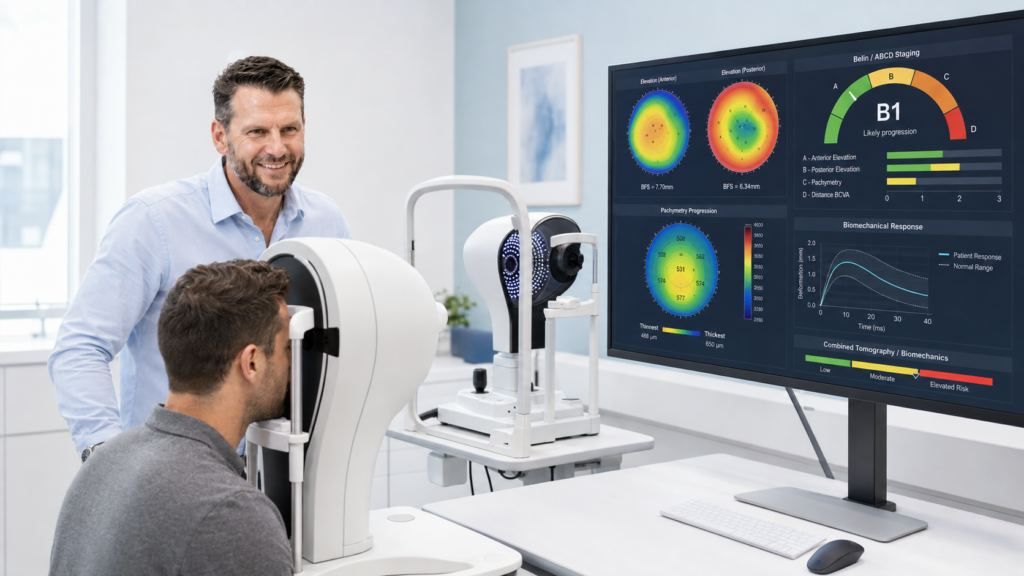
This is why our approach to keratoconus assessment includes both the Pentacam® AXL Wave and Corvis® ST. The Pentacam AXL Wave evaluates corneal shape, including the posterior surface and thickness distribution. The Corvis ST evaluates how the cornea responds dynamically to a controlled air pulse. Together, they allow us to assess both sides of the ectasia question:
Has the cornea’s shape begun to change—and does its mechanical behaviour suggest that it is vulnerable?
Why the back may reveal the problem early
The corneal stroma is not mechanically uniform through its depth.
Laboratory studies by Randleman and colleagues demonstrated a marked depth-dependent variation in cohesive tensile strength. The anterior stroma is substantially stronger, while strength decreases toward the posterior cornea. Petsche and colleagues reported a corresponding depth-dependent gradient in transverse shear properties.
There is a structural explanation. Anterior stromal lamellae are more densely interwoven and have greater branching and transverse connectivity. Posterior lamellae are arranged more parallel to one another, with less interweaving.
Interwoven lamellae are better able to resist interlamellar movement. More parallel lamellae are potentially more susceptible to slippage and deformation when local tissue integrity deteriorates.
This does not prove that keratoconus always begins anatomically in the posterior stroma. Bowman’s layer disruption and anterior stromal abnormalities may also occur early. It does, however, help explain why posterior corneal shape can become diagnostically abnormal before obvious anterior steepening appears.
That distinction matters: posterior elevation may be an early detectable expression of ectasia, even if it is not necessarily the biological origin of the disease.
Keratoconus is focal before it becomes obvious
Early keratoconus should not be imagined as the entire cornea becoming uniformly softer.
Spatially resolved Brillouin studies support a more localised process. Shao and colleagues found that keratoconic corneas demonstrated focal biomechanical weakening around the cone, with Brillouin measurements changing progressively with distance from that region. Importantly, this spatial abnormality was present in mild disease.
A focal weakness can produce a small island of posterior elevation and an abnormal thickness profile before there is enough global anterior steepening to create classic topographic keratoconus.
That is one reason a normal-looking axial curvature map cannot, by itself, exclude early disease.
The anterior surface can disguise the disease
The anterior cornea is not a passive surface.
The epithelium remodels in response to the underlying stromal shape. Over a developing cone, it can become thinner centrally and thicker in a surrounding annulus—the characteristic epithelial “doughnut” described in very-high-frequency ultrasound studies.
This remodelling smooths the front of an already irregular stroma.
The tear film provides another optical smoothing layer, filling subtle surface irregularities. Bowman’s layer may also resist or redistribute anterior deformation during the earlier stages of disease.
The posterior surface has none of these masking mechanisms:
- no epithelium;
- no tear film; and
- no Bowman’s layer.
This makes posterior corneal assessment particularly valuable when the anterior surface still appears deceptively regular.
Why patients may remain asymptomatic
Posterior corneal changes can be optically quiet.
The refractive-index difference at the posterior stroma–aqueous interface is much smaller than the difference at the air–tear interface. Consequently, the posterior surface contributes only a relatively small proportion of total corneal power.
A meaningful posterior shape change may therefore produce little immediate refractive effect. The patient may continue to read well, report no symptoms and demonstrate an apparently stable prescription.
Anterior topography is largely concerned with the surface that contributes most to refraction. Tomography asks a different question: what is the three-dimensional shape and thickness distribution of the entire cornea?
That is why elevation-based tomography can reveal abnormalities that refraction and anterior curvature alone may miss.
What the Pentacam AXL Wave adds

The Pentacam AXL Wave uses rotating Scheimpflug imaging to reconstruct the anterior segment and analyse both anterior and posterior corneal surfaces. Unlike anterior curvature-only assessment, it provides information including:
- posterior elevation;
- anterior and posterior curvature;
- complete pachymetric mapping;
- thickness progression from the thinnest point to the periphery;
- displacement of the thinnest point;
- elevation relative to reference surfaces;
- keratoconus and ectasia-screening displays;
- Belin–Ambrósio relational thickness analysis; and
- Belin ABCD keratoconus staging.
It also integrates tomography with optical biometry, whole-eye wavefront analysis, objective refraction and retroillumination in a single platform.
The clinical value is not one isolated red number. It is the pattern.
A small posterior elevation island may be equivocal alone. A relatively thin cornea may still fall within a population reference range. An anterior curvature map may appear almost normal. When posterior elevation, abnormal pachymetric progression, asymmetry and anterior–posterior imbalance occur together, the case becomes more persuasive.
Tomography allows us to see the cornea as a three-dimensional structure—not merely as a refracting front surface.
Why shape is still only half the story
Tomography measures morphology. It tells us what shape the cornea has assumed.
Keratoconus, however, is fundamentally associated with altered mechanical behaviour. Structural weakening may precede, accompany or progress differently from the geometric changes measured on a static map.
This is where the Corvis ST adds a second diagnostic dimension.
The Corvis ST applies a defined air pulse and uses an ultra-high-speed Scheimpflug camera—capturing more than 4,300 images per second—to record the cornea’s deformation and recovery.
Rather than looking only at thickness or curvature, it evaluates how the cornea moves under load.
Its dynamic corneal response measurements include parameters relating to:
- first and second applanation;
- highest concavity;
- deformation amplitude;
- deformation-amplitude ratios;
- integrated inverse radius;
- stiffness at first applanation;
- corneal thickness; and
- biomechanically corrected intraocular pressure.
The Corvis Biomechanical Index combines deformation-response parameters to assist ectasia detection. When Corvis ST data are integrated with Pentacam tomography, the Tomographic Biomechanical Index combines structural and biomechanical information into a unified ectasia-risk assessment.
The important principle is straightforward:
Pentacam shows us the architecture. Corvis ST challenges that architecture and shows us how it behaves.
Why no single measurement is enough
Posterior elevation is valuable, but it is not infallible.
Scheimpflug systems reconstruct the posterior surface through the anterior cornea. Posterior measurements can be noisier than anterior measurements, and small changes should not automatically be interpreted as progression.
Much of the evidence supporting early posterior elevation is cross-sectional. Studies show that posterior abnormalities can be detected in subclinical and forme-fruste keratoconus, but they do not prove that posterior change universally precedes anterior change in every eye.
Posterior elevation also performs better as part of a multivariable assessment than as an isolated diagnostic test. The relationship between anterior and posterior surfaces, pachymetric distribution and biomechanical behaviour is often more informative than one raw elevation value.
For that reason, we do not diagnose keratoconus from a single colour map or index. We look for concordance across:
- anterior and posterior tomography;
- pachymetric progression;
- elevation patterns;
- anterior–posterior asymmetry;
- corneal biomechanics;
- refraction and visual acuity;
- slit-lamp findings;
- family and ocular history; and
- serial change on the same instrument.
Where available, epithelial thickness mapping can provide another useful layer by demonstrating whether epithelial remodelling is concealing an underlying stromal cone.
The practical advantage of combining Pentacam and Corvis ST
The most difficult keratoconus cases are rarely the obvious cones. They are the patients whose results are almost normal:
- the young patient with increasing astigmatism;
- the eye-rubbing patient with excellent acuity;
- the asymmetric sibling of a patient with keratoconus;
- the borderline refractive-surgery candidate;
- the patient with suspicious pachymetric progression but unremarkable anterior curvature; or
- the eye in which one index is abnormal while the remaining maps appear reassuring.
In these cases, anterior topography alone may answer the wrong question.
The better questions are:
- Is there an abnormal three-dimensional relationship between the anterior surface, posterior surface and corneal thickness distribution?
- Does the cornea deform in a way that is consistent with reduced biomechanical stability?
- Do independent structural and biomechanical measurements point in the same direction?
- Is the pattern stable, or is it changing on repeat measurements?
The Pentacam AXL Wave and Corvis ST are valuable because they approach these questions from different but complementary directions.
Earlier information supports better decisions
Early detection does not mean labelling every unusual cornea as keratoconic. It means recognising risk before obvious anterior steepening and visual deterioration make the diagnosis easy.
That can support:
- safer refractive-surgery screening;
- earlier and more appropriately timed review;
- better counselling about eye rubbing and modifiable risk factors;
- more defensible assessment of progression;
- timely referral for corneal specialist opinion; and
- better-informed decisions about corneal cross-linking.
No device replaces clinical judgement, and no index should be interpreted in isolation. But clinical judgement improves when it is informed by the right measurements.
If we assess only the anterior surface, we may be looking at the part of the cornea most capable of disguising early disease.
The Pentacam AXL Wave helps reveal the unmasked posterior geometry and the full pachymetric profile. The Corvis ST adds the missing functional question: how does this cornea behave when mechanically challenged?
For early keratoconus, seeing shape and biomechanics together is not simply more data. It is a more complete way of understanding the cornea.
Selected references
- OCULUS Pentacam AXL Wave—official product information
- OCULUS Corvis ST—official product information
- Randleman et al. Depth-dependent cohesive tensile strength in human donor corneas
- Petsche et al. Depth-dependent transverse shear properties of the human corneal stroma
- Shao et al. Spatially resolved Brillouin spectroscopy in keratoconus
- Kitazawa et al. Anterior and posterior ratio of corneal surface areas
- Reinstein et al. Corneal epithelial thickness profile in keratoconus
- De Sanctis et al. Posterior corneal elevation measured by Pentacam
- Management of keratoconus: an updated review