The 10 Biggest Mistakes in Orthokeratology Fitting and Patient Management
Orthokeratology works—and, with appropriate patient selection, fitting, education and follow-up, it has a reassuring safety profile. Many disappointing outcomes and serious complications can nevertheless be traced to preventable clinical or patient-management errors.
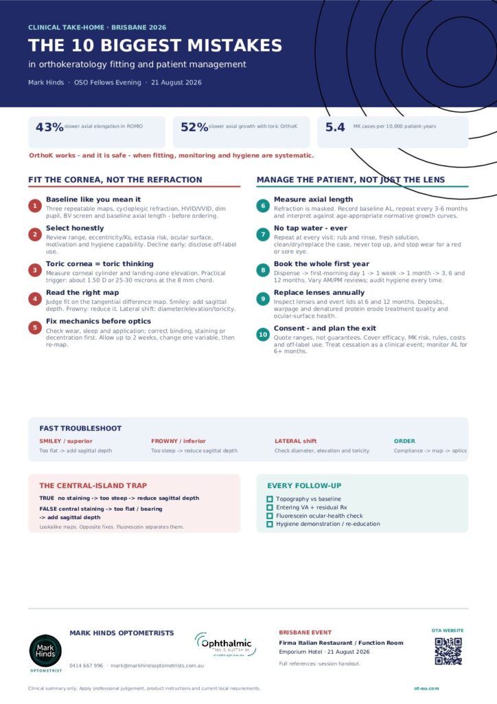
This downloadable clinical handout summarises ten of the most important mistakes encountered in orthokeratology practice and provides practical strategies for avoiding them.
What the guide covers
The guide addresses five common fitting errors:
- Fitting from inadequate baseline data
- Ignoring patient-selection red flags
- Forcing a spherical lens onto a toric cornea
- Misinterpreting corneal topography
- Adjusting the refraction before correcting the mechanical fit
It also examines five patient-management errors:
- Assessing myopia control by refraction rather than axial length
- Providing inadequate hygiene education—particularly about tap-water exposure
- Using an insufficient follow-up schedule
- Failing to establish a regular lens-replacement programme
- Overpromising outcomes, under-consenting patients and poorly managing treatment cessation
Practical orthokeratology troubleshooting
The handout includes a rapid reference for interpreting common treatment-zone patterns, including superior, inferior and lateral decentration.
It also highlights an important clinical trap: true and false central islands can appear similar on a difference map, but their management is opposite. Fluorescein assessment helps distinguish excessive sagittal depth from central bearing caused by an overly flat fit.
Key clinical messages
Reliable orthokeratology begins with repeatable topography, appropriate refraction, corneal and pupil measurements, binocular-vision assessment and baseline axial length.
Mechanical fitting problems should be addressed before optical adjustments are attempted. Hygiene instructions—especially “no tap water, ever”—should be reinforced at every visit rather than discussed only at dispensing.
Axial length, not the morning refraction, is the principal outcome measure when orthokeratology is prescribed for myopia management. Follow-up should be structured, proactive and sustained well beyond the initial fitting period.
Download the clinical handout
Download The 10 Biggest Mistakes in Orthokeratology Fitting and Patient Management for a concise A4 reference suitable for use in clinical practice.
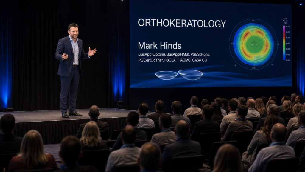
The guide was prepared by Mark Hinds, BScApp(Optom), BScApp(HMS), PGBScHons, PGCertOcTher, FBCLA, FIAOMC, CASA CO, following his presentation at the 2026 Orthokeratology Society of Oceania Fellows Evening in Brisbane.
The complete supporting handout contains the evidence base, references and explanatory notes behind the presentation.
This material is intended for qualified eye-care professionals. It is educational in nature and does not replace individual clinical judgement, applicable product instructions or current professional and regulatory requirements.
Abbreviations used in this session
| AAOMC American Academy of Orthokeratology and Myopia Control | AL Axial length |
| BCLA British Contact Lens Association | BV Binocular vision |
| CLAE Contact Lens and Anterior Eye (journal) | CLEAR Contact Lens Evidence-based Academic Reports (BCLA report series) |
| CASA CO Civil Aviation Safety Authority Credentialled Optometrist | CLS Contact Lens Spectrum (journal) |
| CMN Certified Myopia Management Navigator | DOEE Discontinuation of Orthokeratology on Eyeball Elongation (study) |
| FIAOMC Fellow of the International Academy of Orthokeratology and Myopia Control | GP Gas permeable |
| GPC Giant papillary conjunctivitis | HVID Horizontal visible iris diameter |
| IACMM International Academy Certification in Myopia Management | IAOMC International Academy of Orthokeratology and Myopia Control |
| IMI International Myopia Institute | IOVS Investigative Ophthalmology & Visual Science (journal) |
| LORIC Longitudinal Orthokeratology Research In Children (study) | MK Microbial keratitis |
| OSO Orthokeratology Society of Oceania | OVS Optometry and Vision Science (journal) |
| RCCL Review of Cornea and Contact Lenses (journal) | RCT Randomised controlled trial |
| ROMIO Retardation of Myopia In Orthokeratology (study) | SPK Superficial punctate keratitis |
| TO-SEE Toric Orthokeratology — Slowing Eye Elongation (study) | VA Visual acuity |
Where to get help
- Orthokeratology Society of Oceania — oso.net.au. Education portal, events, mentoring; fellows mentor new practitioners.
- The orthokeratology and myopia control academies — aaomc.org. Certification pathway: Certified Myopia Management Navigator → International Academy Certification in Myopia Management → Fellow of the International Academy of Orthokeratology and Myopia Control.
- BCLA CLEAR – Orthokeratology (refs 4 and 6). The single best free evidence summary to read this month; ref 6 explains what the CLEAR series is.
- GP Lens Institute — gpli.info/corneal-reshaping. Free webinars, troubleshooting guide, clinical pocket guide.
| Mark Hinds BScApp(Optom), BScApp(HMS), PGBScHons, PGCertOcTher, FBCLA, FIAOMC, CASA CO M 0414 667 996 E mark@markhindsoptometrists.com.au Mark Hinds Optometrists / Ophthalmic Trials Australia — Suite 2, 53 Commercial Road, Teneriffe QLD 4005 |
References
Numbered within groups, in the order the evidence appears in the session.
CONSENSUS DOCUMENTS AND EVIDENCE REVIEWS
1. Gifford KL, Richdale K, Kang P, et al. IMI – Clinical Management Guidelines Report. Invest Ophthalmol Vis Sci. 2019;60(3):M184–M203. doi:10.1167/iovs.18-25977
2. Wolffsohn JS, Kollbaum PS, Berntsen DA, et al. IMI – Clinical Myopia Control Trials and Instrumentation Report. Invest Ophthalmol Vis Sci. 2019;60(3):M132–M160. doi:10.1167/iovs.18-25955
3. Flitcroft DI, He M, Jonas JB, et al. IMI – Defining and Classifying Myopia: A Proposed Set of Standards for Clinical and Epidemiologic Studies. Invest Ophthalmol Vis Sci. 2019;60(3):M20–M30. doi:10.1167/iovs.18-25957
4. Vincent SJ, Cho P, Chan KY, et al. BCLA CLEAR – Orthokeratology. Cont Lens Anterior Eye. 2021;44(2):240–269. doi:10.1016/j.clae.2021.02.003
5. Willcox M, Keir N, Maseedupally V, et al. BCLA CLEAR – Contact lens wettability, cleaning, disinfection and interactions with tears. Cont Lens Anterior Eye. 2021;44(2):157–191. doi:10.1016/j.clae.2021.02.004
6. Wolffsohn JS, Jones L, Efron N. CLEAR – Contact lens evidence-based academic reports. Cont Lens Anterior Eye. 2021;44(2):129–131.
CLINICAL TRIALS AND COHORT STUDIES
7. Cho P, Cheung SW. Retardation of Myopia in Orthokeratology (ROMIO) study: a 2-year randomized clinical trial. Invest Ophthalmol Vis Sci. 2012;53(11):7077–7085. doi:10.1167/iovs.12-10565
8. Chen C, Cheung SW, Cho P. Myopia control using toric orthokeratology (TO-SEE study). Invest Ophthalmol Vis Sci. 2013;54(10):6510–6517. doi:10.1167/iovs.13-12527
9. Cho P, Cheung SW, Edwards M. The Longitudinal Orthokeratology Research in Children (LORIC) in Hong Kong: a pilot study on refractive changes and myopic control. Curr Eye Res. 2005;30(1):71–80. doi:10.1080/02713680590907256
10. Cho P, Cheung SW. Discontinuation of Orthokeratology on Eyeball Elongation (DOEE). Cont Lens Anterior Eye. 2017;40(2):82–87. doi:10.1016/j.clae.2016.12.002
11. Chen C, Cheung SW, Cho P. Toric orthokeratology for highly astigmatic children. Optom Vis Sci. 2012;89(6):849–855. doi:10.1097/OPX.0b013e318257c20f
12. Sun L, Li ZX, Chen Y, He ZQ, Song HX. The effect of orthokeratology treatment zone decentration on myopia progression. BMC Ophthalmol. 2022;22(1):76. doi:10.1186/s12886-022-02310-4
SAFETY AND MICROBIAL KERATITIS
13. Hiraoka T, Matsumura S, Hori Y, Kamiya K, Miyata K, Oshika T. Incidence of microbial keratitis associated with overnight orthokeratology: a multicenter collaborative study. Jpn J Ophthalmol. 2025;69(1):139–143. doi:10.1007/s10384-024-01137-4
14. Watt KG, Swarbrick HA. Trends in microbial keratitis associated with orthokeratology. Eye Contact Lens. 2007;33(6 Pt 2):373–377. doi:10.1097/ICL.0b013e318157cd8d
15. Watt K, Swarbrick HA. Microbial keratitis in overnight orthokeratology: review of the first 50 cases. Eye Contact Lens. 2005;31(5):201–208.
16. Bullimore MA, Sinnott LT, Jones-Jordan LA. The risk of microbial keratitis with overnight corneal reshaping lenses. Optom Vis Sci. 2013;90(9):937–944. doi:10.1097/OPX.0b013e31829cac92
17. Kam KW, Yung W, Li GKH, Chen LJ, Young AL. Infectious keratitis and orthokeratology lens use: a systematic review. Infection. 2017;45(6):727–735. doi:10.1007/s15010-017-1023-2
18. Jiang J, Bian Z, Wang F, Lian L, Fan L. Level of compliance in orthokeratology. Eye Contact Lens. 2018;44(5):330–334. doi:10.1097/ICL.0000000000000516
19. Gan C, Ngu K. How can we promote safety in orthokeratology? Myopia Profile; 22 November 2021. myopiaprofile.com/articles/safety-in-orthokeratology
20. Wu J, Xie H. Orthokeratology lens-related Acanthamoeba keratitis: case report and analytical review. J Int Med Res. 2021;49(3):3000605211000985. doi:10.1177/03000605211000985
LENS DEPOSITS AND THE OCULAR SURFACE
21. McCanna DJ, Oh S, Seo J, Coles-Brennan C, Fadli Z, Subbaraman LN, Jones LW. The effect of denatured lysozyme on human corneal epithelial cells. Invest Ophthalmol Vis Sci. 2018;59(5):2006–2014.
22. Omali NB, Subbaraman LN, Coles-Brennan C, Fadli Z, Jones LW. Biological and clinical implications of lysozyme deposition on soft contact lenses. Optom Vis Sci. 2015;92(7):750–757.
23. Messer B. Navigating corneal staining in ortho-k wearers. Contact Lens Spectrum. May 2025.
CLINICAL AND TRADE LITERATURE
24. Simard P, Gagnon F, Michaud L. The most frequent orthokeratology problems to troubleshoot. Review of Cornea and Contact Lenses. January/February 2025:26.
25. Gagnon F, Simard P, Michaud L. Consider these elements when fitting ortho-k candidates. Review of Cornea and Contact Lenses. January/February 2025:20.
26. Michaud L, Gagnon F, Simard P. Orthokeratology principles and design. Review of Cornea and Contact Lenses. January/February 2025:14.
27. Chapman C. Orthokeratology today: topography tells the tale. Contact Lens Spectrum. March/April 2026.
28. Chan K. Ortho-k fitting across the lifespan. Review of Cornea and Contact Lenses. January/February 2026.
29. Chang P. Orthokeratology: principles, lessons, cases. Review of Cornea and Contact Lenses. November/December 2021.
30. Gidosh N. Beyond the basics: ortho-k troubleshooting. Contact Lens Spectrum. June 2024.
31. Potter R. Advanced ortho-k problem-solving. Contact Lens Spectrum. March 2024.
32. Potter R. Orthokeratology for today. Contact Lens Spectrum. October 2020.
33. Frogozo M. Troubleshooting orthokeratology for myopia management. Contact Lens Spectrum. March 2020.
34. Ramdass S. Orthokeratology patient management and follow-up. Ortho-K Knowledge Bank, Review of Myopia Management; 14 March 2023.
PROFESSIONAL BODY GUIDANCE
35. American Academy of Orthokeratology and Myopia Control. Practitioner resources, equipment and certification requirements, and frequently asked questions. aaomc.org
36. Orthokeratology Society of Oceania. Education portal, fellowship requirements and patient guidance. oso.net.au
Notes on the evidence
Where the session simplified something, or where a figure in wide circulation could not be verified against the primary source, it is flagged here. Read these before quoting the numbers yourself.
- Microbial keratitis rates (ref 16). Bullimore 2013 reported 13.9 per 10,000 patient-years in children, 0 in adults, and 7.7 overall — but on only two events, with confidence intervals of 1.7–50.4, 0–31.7 and 0.9–27.8 respectively. The authors caution that overlapping intervals are not evidence of equivalence.
- Decentration and myopia control (ref 12). The “severe is greater than 1.0 mm” grading is Sun 2022’s own. However, that study found no significant difference in axial elongation between decentration groups. The argument that decentration degrades optical quality comes from its discussion section, not its results.
- Rebound after discontinuation (ref 10). The commonly quoted 0.15 mm versus 0.08 mm figures come from secondary sources; the published abstract describes a seven-month discontinuation phase, not six. The authors’ own conclusion — that stopping at or before age 14 led to more rapid axial elongation — is solid and is what the session relies on.
- Time to infection (ref 17). The frequently cited mean of about 19 months of wear before infection reaches the session by way of Gan & Ngu, whose source is Kam 2017. It is not stated in the Kam abstract, so treat it as indicative until confirmed against the full text.
- “20/happy”. A widely used clinical aphorism rather than a sourced claim. It is good advice; it is not a citation.
- Axial length benchmarks (ref 1). The 0.1 mm/yr normal and 0.2–0.3 mm/yr progressing figures are from the IMI Clinical Management Guidelines Report, which states in the same paragraph that there are no established criteria for normal or accelerated axial elongation in a given individual. Use normative growth curves, not a single threshold.
- Toric orthokeratology entry criteria (ref 11). Chen 2012 enrolled children with with-the-rule astigmatism of 1.25–3.50 D. “Corneal cylinder” is a slightly stronger claim than the paper supports.